Biomedical Engineering Reference
In-Depth Information
Ventricular Cell
0
1
0
3
2
0
mV
0
3
mV
−
50
4
−
50
4
4
4
−
100
g
Ca
++
g
K
+
g
K
+
200 ms
Ion
Conductances
Ion
Conductances
g
N
+
g
N
++
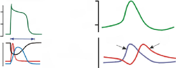
Fig. 6.7
(
Left
) action potential of the ventriculomyocyte (classical model) with its five phases
0-4, phase 4 being the resting membrane potential. Phase 0, the rapid depolarization phase, is due
to opening (influx) of fast Na
+
channels (with quick rise in membrane conductance g
Na
+
, current
INa) and closure of K
+
channels. Phase 1 is due to closure of the fast Na
+
channels and transient
outward fluxes of K
+
and Cl
−
. Phase 2 (plateau) is caused by a balance between Ca
2
+
influx
using long-lasting channels and K
+
outflux. Phase 3, repolarization down to the resting membrane
potential, is induced by a decrease in Ca
2
+
influx and K
+
outflux. During phases 0, 1, 2, and part
of phase 3, the cell is refractory to the initiation of a new action potential. (
Right
) action potential
spontaneously generated (automaticity) in pacemaker cells of the sinoatrial node. A continuous
K
+
outflux causes slow depolarization. Phase 0, fast depolarization due to Ca
2
+
influx. Phase 3,
repolarization, is due to inactivation of voltage-gated Ca
2
+
channels, decaying Na
+
flux, and rising
K
+
flux (from [
761
] (web site) with author permission).
potentials; Fig.
6.7
) is due to a K
+
outflux associated with Na
+
influx and a small
Ca
2
+
influx. Once the depolarization reaches a threshold of about
40 mV, a new
action potential is triggered. Quick depolarization (corresponding to phase 0) is
mainly caused by an augmented Ca
2
+
influx. Repolarization occurs (corresponding
to phase 3) when Ca
2
+
influx decreases and K
+
outflux increases.
Involved mechanisms include the modulating hyperpolarization-activated inward
Na
+
current,
61
inward T- and L-type Ca
2
+
currents, time-dependent decay of
K
+
conductance, Na
+
-Ca
2
+
exchange, and low background K
+
conductance.
Voltage-activated Ca
2
+
release (Ca
2
+
sparks) significantly contributes with the
activity of multiple sarcolemmal ion channels to heart electrical excitations that
originate from the sinoatrial node as well as latent atrial pacemakers. This voltage-
gated intracellular Ca
2
+
release is triggered by the activation of T-type Ca
2
+
channels (Ca
V
3) [
623
]. Atrial pacemaker cells do not contain T tubules. Therefore,
subsarcolemmal cisternae of the sarcoplasmic reticulum are located along the cell
periphery with a subspace (size
−
∼
25 nm) rich in Ca
V
3 channels.
61
Pacemaker cells lacking hyperpolarization-activated inward Na
+
current have a lower frequency.
Search WWH ::

Custom Search