Biomedical Engineering Reference
In-Depth Information
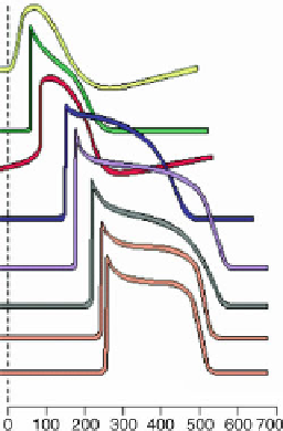
Fig. 6.6
Varying shapes
(time in ms) of the action
potential during its
propagation in the nodal
tissue. From top to bottom:
sinoatrial node, atrial
myocardium, atrioventricular
node, His bundle and its
branches, Purkinje fibers, and
ventricular myocardium
(Source: [
622
]).
Table 6.16.
Nodal tissue and ionic currents (Source: [
468
]). According to the types of involved
ion channels, the initial depolarization ramp (upstroke; V/s) is more or less steep, action potential
duration (APD) more or less long (ms), and firing frequency more or less high (beats/mn). Action
potential duration is longer in the crista terminalis than in pectinate muscles in the right atrium,
and longer in the right auricle than the left atrium. The short action potential in atria is partially
explained by
i
K
,
ur
current. Currents
i
Ca
,
L
and
i
Ca
,
T
are responsible for phase 0 in the sinoatrial node
(SAN) and
i
Ca
,
L
in the atrioventricular node (AVN). Non-selective cation “funny” current
i
f
in the
atrioventricular node is responsible for a secondary pacemaker activity (A: atrium; V: ventricle;
H: His bundle; P: Purkinje fibers; endo: subendocardial; epi: subepicardial; mid: midmyocardial
layers of the myocardium).
Current
SAN
A
AVN
H
P
V
i
K
,
ur
0
+
0
0
0
0
i
K
,
r
+
+
+
+
+
+
i
K
,
s
+
+
<
SAN
+
+
+
i
K
,
to
+
+
+
+
Mid, epi
>
endo
i
K1
∼
0
Low
Low
<
V
<
V
+
i
K
ACh
+
+
+
+
i
K
AT P
+
+
+
+
i
f
+
+
+
+
+
+
i
Na
0
+
+
+
+
+
+
+
<
<
+
i
Ca
,
L
0
V
V
+
+
+
+
+
+
i
Ca
,
T
Frequency
70-80
50
30-60
20-40
15-30
15-30
Upstroke
2-5
150-250
10-20
400-600
500-600
APD
150
150
150
400
400
epi
<
endo
<
mid


Search WWH ::

Custom Search