Biomedical Engineering Reference
In-Depth Information
16 layers
30 nm
θ
2
θ
1
7 nm
25 nm
5 nm
(C)
(A)
(B)
23 layers
10 nm
(D)
(E)
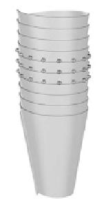
FIGURE 12.7
Microstructure of a scrolled nanotube. (A) TEM image showing a cork-shaped catalyst particle at the tip of a tube,
and the presence of an inclined angle
1
relative to the tube axis. (B) HRTEM images of the tube at a marked loca-
tion showing interruption of lattice fringes only at external walls and a reduction of layer number (Note:
1
2
).
(C) HRTEM image of the base tube showing the interruption of the external and internal lattice fringes of the nano-
tube wall and an inclined angle of
2
with the tube axis. (D) Decoration of Pt nanoparticles revealing a preferential
path along the edges of scrolled structure. (E). 3D model of the scrolled structure and deposited Pt nanoparticles
on the edges. (From Sun, X., Li, R., Stansfield, B., Dodelet, J. P., Ménaed, G., Désilets, S., Unpublished work)
on the surface of the tubes. After a slight oxidation treatment of the tubes [42], a preferen-
tial deposition of Pt NPs (79 among 112 particles counted on the micrograph in Figure
12.7D) are aligned along the spiraling external edge of the graphene sheet, as shown
schematically by the 3D model in Figure 12.7E. Actually, the scroll growth model had been
previously proposed for nanofibers [43] and observed in hydrothermal synthesis [44].
Similar to SWCNTs, to further elucidate their growth mechanism, it is essential to under-
stand how nanometer catalyst particles initiate the growth of MWCNTs. So far, there have
been few reports and little direct evidence concerning the initiation of the growth of nan-
otubes due to the extremely fast growth process. Sun et al. [41] observed nanotubes at var-
ious stages during their growth by HRTEM measurements of tubes grown from catalyst
particles deposited on MWCNTs (see Figure 12.8). Clearly, the initial shape of the catalyst
particles deposited on the nanotubes is spherical before growth begins, with a diameter of
about 35 nm (Figure 12.8A). In the early stages, the catalyst particle was reshaped to reveal
faceting (Figure 12.8B). It can be seen clearly that a thicker graphitic wall appears at the bot-
tom of the catalyst particle, indicating that the initiation of the structure occurs from the