Biomedical Engineering Reference
In-Depth Information
Essentially, the bioengineered interfaces will facilitate (a) characterization of the quanti-
tative boundary conditions of the human body; (b) determination of variation of those
conditions with disease and environmental change; and (c) putting into effect suitable
therapeutic response, as needed.
10.3
Anatomy of Skin
To develop a long-lasting interface, the nuances involved in interfacing with skin need to
be understood. Toward that goal, a brief review of the skin is discussed below.
10.3.1
General Structure
Skin is the largest organ of the human body and accounts for
16% of the body weight. It
serves multiple purposes, which can be broadly classified into four functions—(1) Protection:
physical, biological, against UV light, and from dehydration; (2) Thermoregulation: control of
body temperature; (3) Metabolic functions: synthesis of vitamin D, excretion of waste prod-
ucts, and storage of fat; and (4) Sensory functions: pressure, heat, cold, contact, pain, as well
as integral interaction with vision (pigmentation patterns) and smell (pheromone release). It
is evident from its range of functionality that the skin hosts complex physiological, biome-
chanical, and biochemical processes, all occurring simultaneously.
The skin possesses a layered structure comprising various kinds of tissues. It icomprises
two layers: the dermis and the epidermis. The epidermis is the outer layer of the skin.
Being a tough and waterproof layer, it protects the inner regions. The underlying dermis
is a thicker layer and is responsible for imparting strength and elasticity to the skin.
Beneath the dermis is the subcutaneous layer, which is not part of the skin itself. It is a
layer of tissue made of protein fibers and adipose tissue (fat). It contains glands, sensory
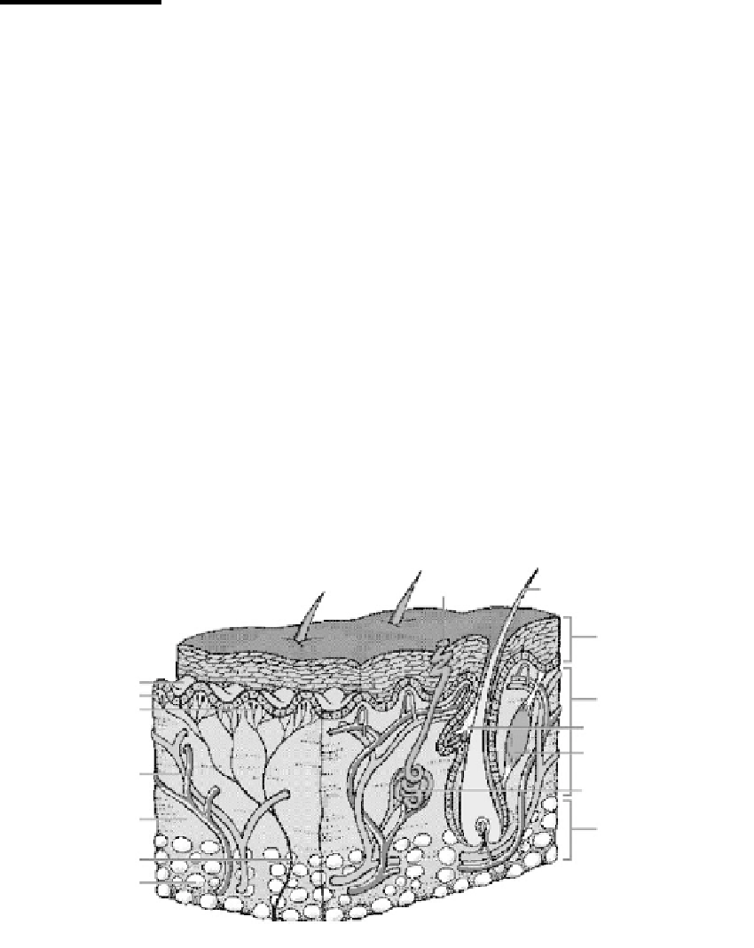
receptors, and other skin structures. Figure 10.3 depicts the cross-section of the skin [1].
Pore
Hair
Epidermis
Dermal papillae
Cold receptor
Heat receptor
Dermis
Sebaceous gland
Arrector pili muscle
Blood vessels
Sweat gland
Connective tissue
Subcutaneous layer
Nerve
Fat lobules
FIGURE 10.3
Cross-section of skin [1]. (From “Structure of the Skin”
Microsoft
®
Encarta
®
Encyclopedia
. http://encarta.msn.com©
1993
-
2004 Microsoft Corporation. All rights reserved, With permission.)


