Biology Reference
In-Depth Information
A
B
C
D
E
F
F
H
H
G
G
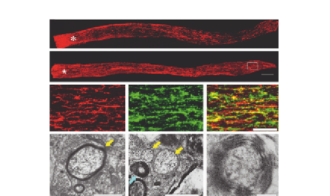
Figure 6.3 Long-distance axon regeneration. Combinatorial treatment enables axons
to grow through the entire length of the optic nerve. Regenerating axons are visualized
by anterograde staining with cholera toxin B fragment (CTB) staining 6 weeks (A) and
10 weeks (B) after injury. (C
-
E) Distal part of the optic nerve coimmunostained for CTB
(red, C), GAP-43 (green, D), and merged images (E). (F
-
H) Electron micrographs of cross-
sections through regenerating optic nerves show fiber with thin myelin (yellow
arrow, F), unmyelinated axons (yellow arrows, G), and a fiber with multiple lamellae of
tightly packed myelin (blue arrow, G) at a higher magnification. Scale bars: (A, B),
200 mm; (C
-
E), 50 mm; (F, G), 1 mm; (G), 0.1 mm.
lateral geniculate nucleus and superior colliculus, in entraining daily activity
patterns with the ambient dark-light cycle (suprachiasmatic nucleus), pupil-
lary constriction (olivary pretectal nucleus), and stabilization of the visual im-
age to compensate for self-generated movements (medial terminal nucleus).
Most mice that received the combinatorial treatment showed at least some
reinnervation of these areas, and a few showed moderately dense rein-
nervation (
de Lima et al., 2012
;
Fig. 6.4
). However, very few axons were
found in inappropriate brain areas. Another study has examined the effects
of deleting the
pten
and
soc3
genes while applying CNTF, and found that this
combination also profoundly increased the number of axons regenerating
down the mouse optic nerve compared to any of the treatments alone, but
the numbers of axons dropped continuously along the length of the optic
nerve and almost none crossed the chiasm (
Sun et al., 2011
). A third instance
of combinatorial treatment has involved overexpression of the histone
acetyltransferase P300 while inducing intraocular inflammation. Together,
these treatments have an additive effect (
Gaub et al., 2011
).