Biology Reference
In-Depth Information
Neu-N
+
CTB
A
B
OPT
MTN
C D E F G H
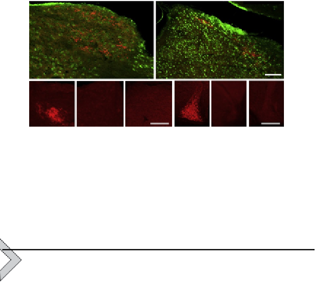
Figure 6.4 Reinnervation of visual nuclei. Reinnervation of central visual targets
10 weeks after optic nerve injury, including the dorsal lateral geniculate nucleus
(dLGN, A) and superior colliculus (SC, B), bothofwhichare stained for CTB (red) to visualize
regenerating axons and Neu-N (green). Regenerating axon terminals stained for CTB in
theolivarypretectal nucleus (OPT, C) andmedial terminal nucleus (MTN, F). No fiberswere
seen on the side ipsilateral to the regenerating optic nerve (D and G) or in control animals
with incomplete regeneration (E and H).
6. RGC SURVIVAL
As noted earlier, increasing cell survival does not by itself lead to axon
regeneration, although it is obviously a prerequisite for regeneration to oc-
cur. RGCs begin to die a few days after intraorbital optic nerve damage and
fewer than 5% remain after a month (
Berkelaar, Clarke, Wang, Bray, &
Aguayo, 1994
). The mechanisms responsible for RGC death are not entirely
clear, and most strategies aimed at preventing cell death are only partially
successful (
Kanamori, Catrinescu, Kanamori, et al., 2010; Kermer,
Klocker, & Bahr, 1999; Koeberle & Ball, 1999; Malik, Shevtsova,
Bahr, & Kugler, 2005; Monnier et al., 2011
). One early event is
activation of the unfolded protein response, and blocking this response
can provide substantial protection but without enhancing regeneration
(
Hu et al., 2012
). Caspases-3, -6, -8, and -9 are all upregulated after optic
nerve injury, but inhibiting these provides only transient protection and
RGCs continue to die at a slower pace (
Berkelaar et al., 1994; Kermer
et al., 1999; Monnier et al., 2011
). Levels of the antiapoptotic proteins
bcl-2 and bcl-x
L
decrease after optic nerve injury (
Isenmann, Wahl,
Krajewski, Reed, & Bahr, 1997; Levin, Schlamp, Spieldoch, Geszvain, &
Nickells, 1997
), and overexpressing either of these, or preventing an