Biomedical Engineering Reference
In-Depth Information
d
n
4
t
3
n
g
|
7
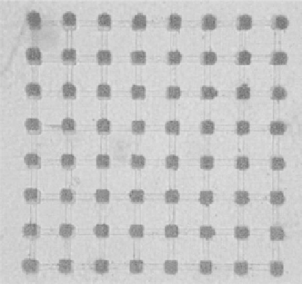
Figure 3.9
A microelectrode array showing planar 50 mm
2
electrodes (Panasonic
Medprobe).
n
3
.
high resolution recording channels, operational amplifiers, filters and long-term
data acquisition instrumentation. The experiments also employ electrical
stimulation systems, real-time signal visualization and optical surveillance of
the cell development using a TV camera connected to an optical microscope
positioned over the array. Parameters of interest such as the time of a burst
occurrence, the time duration between the first and last spikes, the interval
between bursts, changes in synchronization, and the burst amplitude are all
recorded and subjected to subsequent analysis.
The analysis of the spatio-temporal patterns in the activity of the neural
network provides an insight into the internal dynamics of the neural networks,
as well as the relation of synchronization changes to the action of neuro-
transmitters and their blockers.
6
Figure 3.10 shows a hybrid neuron-MEA
interface integrated within a system for neuroscience research aimed towards
the understanding of memory formation and application to artificial
intelligence.
44
Hybrid biosensing systems require both the attachment of neurons to a
substrate surface and the complete preservation of the neuron's biological
activity, viability and functionality upon attachment. In contrast to the
patch clamp technique, such systems are not invasive and are capable of
monitoring the changes in cell metabolism as a result of input stimuli. The non-
biological surface has to be sterile and hospitable to support a neuron or cell. In
order to achieve this, the surface is often modified with organic molecules
recognizable by the cultured cells. Cell attachment can take place by physi-
sorption, chemisorption or by trapping the neurons in polymeric matrices. Care
must be exercised to prevent the creation of unnecessary diffusion barriers that