Biology Reference
In-Depth Information
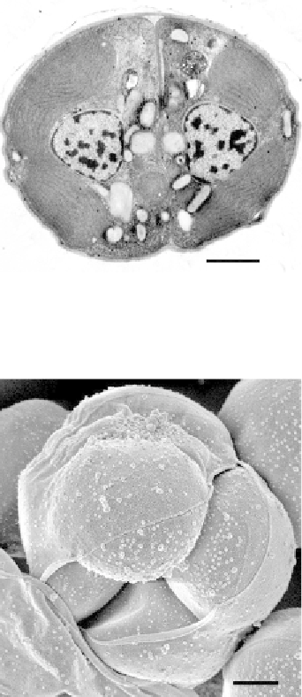
Figure 8.4 Ultrastructure of the asexual cell division of C. velia. The longitudinally sec-
tioned plastids are filled with thylakoids arranged in stacks of three. The autospores also
contain an oval nucleus with nucleoli, as well as numerous granules likely with amylo-
pectin and lipid contents (scale bar¼2
m
m).
Figure 8.5 Detailed view of an autosporangium of C. velia. Ruptured wall reveals the
presence of four tightly bound autospores (scale bar
¼
1
m).
m
Along with the nonmotile cells, elongated zoospores possessing two
prominent flagella (
Fig. 8.8
) and retaining a relatively large and evenly
pigmented plastid (
Fig. 8.9
) were observed to move very fast in a character-
istic zig-zag manner (
Oborn´k et al., 2011; Weatherby et al., 2011
). Zoo-
spores of chromerids highly resemble colpodellids in morphology, overall
cell shape, and the presence of two heterodynamic flagella, both thinly
tapered at their terminus (
Leander et al., 2003
). The motile stages display

Search WWH ::

Custom Search