Biology Reference
In-Depth Information
Figure 8.6 Cortical alveoli below the plasma membrane of C. velia. Section through the
periphery of a vegetative cell reveals the presence of flat electron-dense alveoli and a
single layer of underlying microtubules (scale bar¼200 nm).
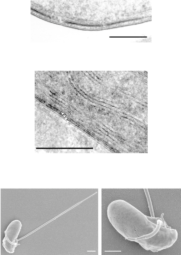
Figure 8.7 Membranes of the C. velia plastid. Arrowheads point to four membranes sur-
rounding this secondary plastid. Note the arrangement of thylakoids in stacks of three
(scale bar¼100 nm).
Figure 8.8 Scanning electron microscopy of C. velia flagellated zoospores. The elon-
gated zoospores are equipped with two heterodynamic flagella. The shorter flagellum
contains a typical finger-like projection (scale bar¼1
m
m). The left panel is reprinted from
Oborník et al. (2011)
, Copyright (2011), with permission from Elsevier 2011.

Search WWH ::

Custom Search