Biomedical Engineering Reference
In-Depth Information
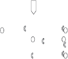
(a)
(c)
OH
-
Acetic
Collagen
(b)
(d)
nano-HA
PO
4
-
Ca
2+
Figure 16.2.
Biomimetic self-assembly method of producing nano-HA-collagen composite.
(A): Type I collagen was dissolved in acetic acid. (B): Ca
2+
and
PO
−
were added. (C) Drops of
NaOH were incorporated into the solution for the co-precipitation of calcium phosphates with
collagen. (D) Aging the precipitates for more than two hours. (E): Centrifugation of solution
for the retrieval of nano-HA (not shown)
71
(Adapted from
Nanomedicine
(2006) 1(2), 177-188
with permission of Future Medicine Ltd.)
The nanoHA - collagen nanocomposite has nano - sized, bone - like apatite
embedded in the collagen matrix. The three hierarchical levels—namely the calci-
fi ed collagen fi brils, collagen molecules, and fi bers—showcase an example of a
self-assembly biomimetic material, where the crystallographic axes (
c
- axes) of
the nano-HA crystals are intimately aligned with the longitudinal axes of the col-
lagen fi brils. The nano-HA and collagen molecules co-precipitated into mineral-
ized collagen fi brils are approximately 6 nm in diameter and 300 nm in length as
shown in Figure 16.3
73
. The presence of this bone-like mineral is one of the pre-
requisites of good interfacial bonding with the orthopedic implants with the host's
bone (i.e. osteoconductivity) and may trigger osteogenic differentiation of pro-
genitor or bone cells (i.e. osteoinductivity). Table 16.3 summarizes the various
types of nanocomposites and their respective cellular responses
70,72,74 - 86
.
Although cell culture results were promising where osteoblasts adhered to
the biomimetic nano-HA/collagen/PLA scaffold within two days of culture, sub-
sequently proliferated within the pores of the materials and within a week, full
confl uence was achieved, the material was tested in an
in vivo
setting to evaluate
its effi ciency as a potential bone graft material
73
. A 15 mm segmental defect was


Search WWH ::

Custom Search