Biomedical Engineering Reference
In-Depth Information
Y
X
Z
FIgurE 13.4
(
See color insert
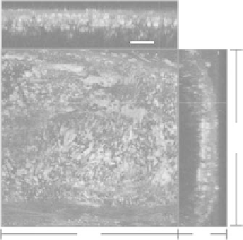
.) Atherosclerotic lesion in a ligated mouse carotid artery. Elastin autofluo-
rescence is green, lipid droplets labeled with Nile Red is yellowish green, and collagen SHG is red. Scale bar:
40 μm. (Reproduced from Yu, W. M. et al. 2007.
J Biomed Opt
12. With the permission of Lippincott Williams
& Wilkins.)
lesions are induced by ligation of the carotid artery in apolipoprotein E-deficient mice 2 weeks prior
to imaging. The induced lesions had increased collagen content and lipid deposition as shown in
Figure 13.4 (collagen (SHG, red), elastin (two-photon excitation fluorescence, green), and lipid drop-
lets labeled with Nile Red (TPEF, yellow)). As in the
in vitro
perfused preparations [32], imaging the
clinically interesting intimal surface requires imaging through the adventitia, limiting the approach
to small arteries (see Figure 13.5 and Section 13.4 for a description of the vessel wall layer). Moreover,
blood flowing through the vessel completely eliminates any possibility of observing the opposing wall.
Although these constrained preparations have provided new insights, it is desirable to observe unper-
turbed tissue
in vivo
.
Dynamic microscopy is an emerging technology that allows imaging of tissue
in vivo
in its natural
undisturbed state. This method can reduce blur or distortion-induced motion by (i) maximizing the
acquisition rate, (ii) synchronizing the image trigger with the respiratory and cardiac cycles [35], and
(iii) fast tracking of image features, using closed-loop schemes for stage control [4]. Such schemes rely on
a metric of displacement, such as the normalized cross correlation, between a reference image/Z-stack
and the updated image/Z-stack. Real-time tracking can be achieved with the use of a dedicated graph-
ics processing unit. Motion-corrected
in vivo
images of mouse tibialis anterior muscle and associated
capillaries were recently obtained using a motion tracking stage in free-breathing anesthetized mice.
The lower leg was immobilized by clamping above the foot without obstructing proximal blood flow
and the skin and surface fascia over the muscle were removed. Two channel images were recorded and
consisted of intrinsic muscle fiber NAD(P)H fluorescence and the dye di-8-ANEPPS to visualize vascu-
lature endothelium, centered at 460 and 590 nm, respectively. The spatial information in the vascular
channel was used for tracking 3D motion on the order of 20 μm/min and increased image sharpness
was clearly demonstrated in both channels [4]. With cyclic triggering, data are only collected during
short acquisition times periodically. An improvement over cyclic triggering is adaptive motion filter-
ing wherein the image is collected with continuous, adaptive precompensation for the periodic motion.
The pseudoperiodic motion is first analyzed via conventional tracking. Once learned, compensation is
applied and regularly updated. The coupling of a dynamic microscope with adaptive filtering to com-
pensate for the repetitive motion associated with the pulse pressure and respiratory motion may over-
come this major limitation in the future.