Biomedical Engineering Reference
In-Depth Information
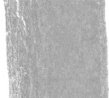
A
M
I
FIgurE 13.5
(
See color insert
.) Structure of the porcine arterial wall. I, intima; M, media; A, adventitia. 25×
magnification, scale bar = 400 μm.
13.4 investigation of Macromolecular Structure
The arterial wall is composed of three layers: the intima, media, and adventitia (see Figure 13.5). The
intima consists of (i) a layer of thin endothelial cells lining the lumen of the vessel and its underlying base-
ment membrane, below which is (ii) a layer of connective tissue containing fibroblasts, macrophages, and
lymphocytes embedded in extracellular matrix. The major components of the extracellular matrix (ECM)
include collagen, elastin, and proteoglycans. The medial layer of the arterial wall is rich in elastic fibers and
smooth muscle cells. The adventitia is composed of collagen and elastic fibers. The intima and media are
separated by a thick layer of elastic fibers called the internal elastic lamina. Similarly, the media and adven-
titia are separated by the external elastic lamina, which is also composed of elastic fibers. The macromo-
lecular structure of collagen, elastin, and proteoglycans determines arterial wall mechanical properties
and susceptibility to pathological remodeling such as atherosclerosis. In the sections that follow, we will
review the advances that SHG and other nonlinear optical imaging technologies have made in our under-
standing of the role that macromolecular components of the arterial vascular bed play in these processes.
13.4.1 elastin
Elastin fibers assemble to form layers that provide long-range deformability and passive recoil to arteries
and are the major determinant of vascular resilience. Elastin fibers are composed of an amorphous core
of the hydrophobic protein elastin surrounded by a mantle of fibrillin-rich microfibrils. Elastin amino
acids are nonpolar (60%), and lysine-residue-linked pyridinoline groups form covalent cross-links
between the chains. Pyridinoline groups exhibit ~400 nm emission maximum when excited in the UV
[37] and may be responsible for elastin two-photon excitation fluorescence [38]. Elastin powder exhibits
an emission maximum at ~480 nm that is also seen in spectra obtained from elastin fibers in human
skin and may correspond to pyridinoline aggregates [38]. Kwon et al. reported that fluorescence emis-
sion of elastin shifted with excitation frequency, suggesting that multiple chromophores are responsible
for elastin fluorescence emission [39].
Optical signal intensities from collagen and elastin can be optimized and spectrally separated (see
Figure 13.6) using an appropriate excitation wavelength and bandpass filters for detection [29,39-42].
An excitation wavelength of 840-860 nm appears to be ideal insofar as it (i) is above the lower end of
SHG detection, (ii) is at the upper limit of elastin detection, (iii) optimizes depth of penetration in the
tissue, and (iv) reduces photodamage due to adsorption.