Biomedical Engineering Reference
In-Depth Information
Normal
Edematous
y
y
y
y
Anterior
stroma
x
x
x
x
50 μm
(a)
(b)
(c)
(d)
z
z
z
z
Posterior
stroma
50 μm
(e)
(f )
(g)
(h)
x
x
x
x
FWSHG
BWSHG
FWSHG
BWSHG
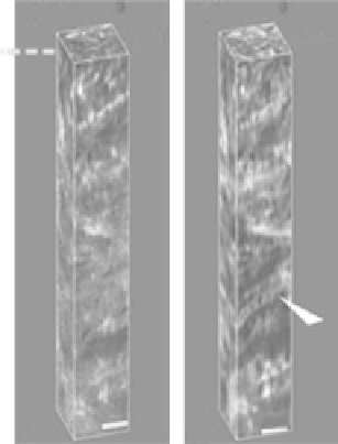
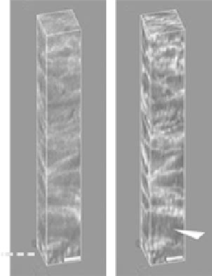
FIgurE 12.12
(a), (b), (e), and (f ) are forward SHG (FWSHG) and backward SHG (BWSHG) image stacks
of untreated bovine corneas. (c), (d), (g), and (h) have been treated with de-ionized water immersion for 2 h
to simulate corneal edema. In both cases, SHG images were acquired from both the anterior and posterior
stroma. Note that in edematous corneas, lamella oriented obliquely to the corneal surface (arrowheads in (d))
and that increased lamellae spacing (arrowheads in (h)) can be visualized. (Adapted from Hsueh, C. M. et al.
2009.
Biophys J
97(4):1198-1205.)