Biomedical Engineering Reference
In-Depth Information
(a)
(b)
(c)
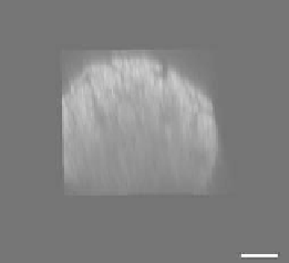
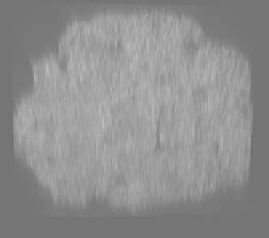
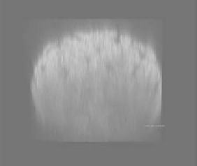
FIgurE 8.8
Optical clearing in the tendon with 50% glycerol is reversible. The 3-D renderings are for the SHG of
(a) uncleared tendon, (b) cleared tendon, and (c) tendon following washing in PBS. Scale bar = 20 μm. (Reproduced
from LaComb, R. et al. 2008.
J. Biomed. Opt.
13:021108.)
cleared collagenous tissues proves that the dipoles are still arranged noncentrosymmetrically in optically
cleared collagen triple helices. Here, we note that no similar arguments have been raised in regard to the
mechanism of optical clearing in the muscle although the collagenous layers of perimysium present in the
muscle are subject to the same considerations as described above. However, the clearing in the muscle is
not likely to be reversible as the cell membranes are permeabilized in the glycerol-cytoplasm exchange.
8.6 optical clearing in Skin
Owing to their regularity and highly organized assembly, striated muscle and tendon were useful exam-
ples in which we can examine optical clearing in depth. We now turn our attention to the more chal-
lenging imaging environment of the skin. Skin is a far more complex collagenous tissue where optical
clearing could offer immediate benefits, as it is readily accessible in clinics. In particular, the dermis is
of significant interest due to the wide range of skin pathologies. This structure is a 1-5 mm thick layer
sandwiched between the epidermis on top and subcutaneous tissue below and is composed of collagen,
elastin, and fibroblasts.
The clearing of the human skin
ex vivo
with DMSO has been shown to introduce morphological
changes to the collagen matrix on a submicrometer scale in addition to refractive index matching [39].
Obviously, the reduction of scattering at the price of decomposition of higher-order collagen structures
limits the use of DMSO only to
ex vivo
imaging. Glycerol does not exhibit such toxicity, especially at
moderate concentrations, since the clearing is reversible as discussed above, and might be a viable agent
for
in vivo
imaging.
One of the most recent studies of
in vivo
optical clearing was carried out on the skin, where 20%, 30%,
and 75% glycerol was injected in the dermis of rat dorsal skin, followed by recording of reflectance spec-
tra
in vivo
, although SHG images were taken from excised samples [19]. A simple reflectance measure-
ment only 10 min after the injection showed stronger reduction of scattering consistent with increasing
concentration of glycerol (see Figure 8.9). For lower concentrations, reflectance recovered and exceeded
its initial level within 30 min time frame, and samples swelled due to the hyperosmotic effect. However,
in the case of 75% glycerol, reflectance remained very low and the samples became thinner instead of
swelling, consistent with almost a 30% decrease in average fibril diameter as measured with electron
microscopy (from 109.34 ± 20 to 79.47 ± 13 nm). The thinning of fibrils can be explained by two-step
dehydration: first the extracellular matrix is dehydrated, creating a gradient of liquid between the fibers
and their surroundings, and then the fibers are dehydrated as well. Neither dissociation of collagen nor
any deterioration of fibers was observed in SHG images, and the signal intensity remained strong with
all three concentrations. In addition to refractive index matching, more regular packing of collagen
fibrils is thought to facilitate optical clearing in the skin [19].