Biomedical Engineering Reference
In-Depth Information
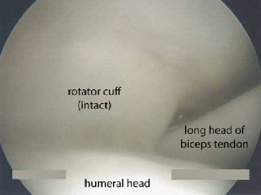
Fig. 12.2
The rotator cuff cable is a relatively hypovascular area near the insertion of the rotator
cuff. Most tears arise in this area, which can be best appreciated from the articular side of the
tendon
area can be visualized from the articular side of the tendon. There is a thickening of
the supraspinatus that forms a cable-like structure, within which is the hypovascular
zone. Tears of the rotator cuff often originate in this zone.
The insertion site of the rotator cuff to bone is comprised of four zones;
this transition provides a gradual transition from a compliant to a hard material [
8
,
9
]. The zones include tendon, unmineralized fibrocartilage, mineralized
fibrocartilage, and bone (Fig.
12.1
). There is a distinctive mineral gradient across
the zones, transitioning from completely unmineralized tissue in the tendon to
highly mineralized tissue in the bone. The purpose of this relatively gradual transi-
tion is to reduce the high stresses that would otherwise concentrate at the junction of
a compliant material (tendon) to a stiff material (bone) [
10
]. These high stresses may
otherwise result in a tear or attenuation of the tendon. The fibrocartilaginous
transition zone may therefore serve a protective role to the tendon as it inserts on
the tuberosity, allowing effective load transfer from the muscles to the bone.
12.3 Biology of Rotator Cuff Tendon-to-Bone Healing
Rotator cuff healing is characterized by a reparative rather than a regenerative
response [
11
,
12
]. A disorganized fibrous interface develops at the insertion
consisting of biomechanically inferior tissue (Fig.
12.3
). This tissue is later
remodeled, becoming more organized and able to bear higher loads. However,
the tissue never
remodels
to entirely resemble normal uninjured tendon.
Search WWH ::

Custom Search