Biomedical Engineering Reference
In-Depth Information
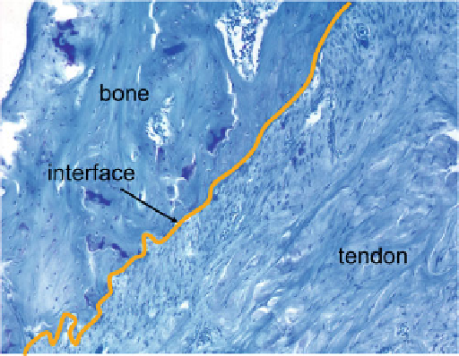
Fig. 12.3
A 4-week specimen after a rotator cuff injury and repair demonstrates relatively
disorganized deposition of extracellular matrix components. Repair tissue is hypervascular and
hypercellular compared to normal tendon tissue. Toluidine blue stained section,
solid line
indicates
interface between healing tendon and bone (reproduced, with permission, from ref. [
12
])
Histologically, the resultant tissue is hypercellular and hypervascular relative to
uninjured tissue. Biomechanical properties likewise never reach those of uninjured
tendon.
Tendon healing occurs in phases similar to those seen in typical wound healing
scenarios. An initial inflammatory phase is characterized by the presence of multi-
nucleated cells. Most of these cells are replaced by mononuclear cells, which are the
predominant cell type present for the next few weeks. In an acute repair rodent
rotator cuff model, metabolic activity, growth factor production, and cell prolifera-
tion reach a peak at 7-10 days [
11
], after which there was a gradual decrease to
baseline levels. While this timing may differ for repairs of chronic tears, and for
healing in humans, there is likely a short time period of cell proliferation and growth
factor production; this timing may provide a window for therapeutic manipulation.
The next phase of tendon healing consists of extracellular matrix production.
While normal tendon is comprised primarily of collagen type I, the healing
response is characterized by an initial production of collagen type III. Collagen
type III is typically seen in degenerated and injured tendon, and also in developing
tendon. It is a primary component of the early fibrous interface at the healing
tendon-to-bone junction. Collagen type I production begins at some point after
collagen type III and persists throughout the course of tendon healing.
Many different growth factors have been isolated at the healing rotator cuff
tendon-to-bone insertion [
12
,
13
]. These include TGF-
, PDGF-BB, bFGF, and
various bone morphogenetic proteins (BMPs). These factors and many others likely
play significant roles in the biology of tendon healing. The exact roles they exert
remain a subject of continuing investigation.
b
Search WWH ::

Custom Search