Biomedical Engineering Reference
In-Depth Information
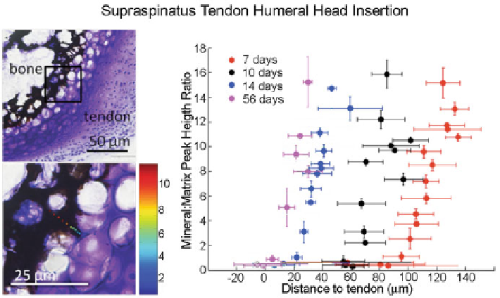
Fig. 11.6
Spatial gradients in mineral (as determined using Raman spectroscopy) form between
tendon and bone at the developing entheses from the onset of endochondral ossification (7 days in
the mouse supraspinatus tendon-to-bone insertion, shown here)
type II collagen, characteristic of cartilage. Neonatal collagen II gene expression was
confirmed by in situ hybridization. The tendon side of the insertion expressed type I
collagen, which is characteristic of tendon and bone ECM. As mineralization of the
secondary ossification center proceeded over the first few weeks of postnatal
development, expression of collagen II became restricted to a narrow zone in the
transitional tissue of the insertion. Type I collagen was expressed by cells adjacent to
the band of collagen II expressing cells on the tendon side of the insertion.
Type X collagen was evident in a band of cells on the bone side of the insertion,
adjacent to the mineralizing front. Collagen X is typically localized to hypertrophic
chondrocytes in the growth plate prior to mineralization. At the enthesis, expression
of this marker persists after the large hypertrophic cells associated with mineraliza-
tion are no longer seen, suggesting that this molecule may play a role in maintaining
the mineralized interface. This result is consistent with the development of the rat
Achilles tendon [
107
].
As previously mentioned, the appearance of a mineral gradient in the transitional
tissue of the insertion likely plays an important role in mediating the transfer of muscle
loads from soft tissues to bone. In the mouse, a mineral gradient is evident near the
developing insertion as early as 1 week after birth (Fig.
11.6
). The mineral gradient
coincides with the mineralizing front of the secondary ossification center in the
humeral head. The mineral gradient is first separated from the developing tendon by
a region of epiphyseal cartilage yet to be mineralized. In a murine model, the gradient
gradually moves into the developing transitional tissue of the tendon-bone insertion as
the epiphyseal cartilage is mineralized between the first 2 weeks of postnatal growth.
Search WWH ::

Custom Search