Biomedical Engineering Reference
In-Depth Information
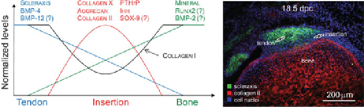
Fig. 11.7
Spatially and temporally controlled expression of a number of transcription factors,
growth factors, and transcription factors likely play important roles in enthesis development
11.4.2 Biological Factors Necessary for Insertion Development
Development of the complex transitional tissue found at the tendon-to-bone
insertion requires precise spatial and temporal control of a range of biological factors
(Fig.
11.7
). The development of transitional tissue occurs during postnatal develop-
ment when tendon and bone are already established, but still actively growing and
remodeling. In order for transitional tissue to develop at the interface, there must be
concurrent regulation of and interaction between the biological signals of tendon,
bone, and cartilage. Individually, all three tissue types are responsive to the mechan-
ical environment. Specific biological molecules native to each tissue type have been
implicated in the cellular response to the mechanical loading environment.
The mineralized side of the insertion is not yet mature bone during the early
stages of enthesis development. Instead, it is an immature epiphyseal cartilage
template undergoing endochondral ossification. At this stage of development, the
insertion shares many features with the growth plate. Biological factors identified at
precise spatial locations in the growth plate include: PTHrP, Ihh, Ptc, Sox9, and
type X collagen [
20
,
112
]. Chemical gradients of these molecules are responsible
for maintaining the graded morphology of the growth plate. These factors have also
been localized to the developing tendon-to-bone insertion and may also impact
development of a graded insertion [
107
-
110
,
113
].
PTHrP was originally described for its role in regulating the growth plate in a
negative feedback loop with Ihh [
81
-
83
]. Recently, PTHrP has been localized
to tendon and ligament entheses during postnatal development [
87
,
113
]. More
specifically, it is localized to a group of fibroblast-like cells in the intermediate zone
between the tendon proper and the transitional tissue that inserts into the underlying
cortical bone [
113
]. Furthermore, PTHrP has been generally localized to periosteal
cells in addition to cells that will form the secondary ossification center of long
bones [
113
]. Elevated expression of PTHrP at tendon-to-bone insertions suggests
that PTHrP may be important to maintain the mineralized interface during devel-
opment. In the growth plate, PTHrP maintains chondrocyte proliferation and blocks
maturation and mineralization [
20
]. PTHrP may have a similar function for enthesis
development.
Search WWH ::

Custom Search