Biomedical Engineering Reference
In-Depth Information
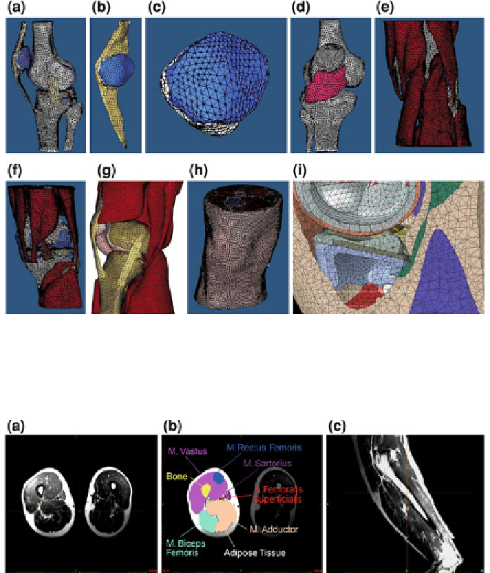
Fig. 5.61 Detailed FE-model of the human knee: a collateral ligaments (yellow), b) detail of
ligament patellae (yellow) and patella (blue), c patella, d H
OFFA
's fat pad (magenta); muscle
groups: e flexors, f rotators, g extensors; h knee with skin and adipose tissue, i in detail: tibia-
femoris-roll and slide process
Fig. 5.62 a Transversal MR-scans of the right and left upper leg, and b reconstruction shading of
anatomical structures (bone, adipose tissue and muscle groups), c sagittal MR-scan
view of the tibia-femoris-roll and slide motion. H
OFFA
's fat pad is located in the
knee joint between the tibia heads (condylus tibiae), the ligament of the patella
(ligamentum patellae) and the lower edge of the patella and is covered with a joint
inner skin (membrana synovialis) composed of loose connective tissue including
synovial cells which provide lubrication. Figure
5.61
h shows the finite element
model of the complete knee region and Fig.
5.61
i shows the anatomical structures
involved in the tibia-femoris-slide motion.