Biomedical Engineering Reference
In-Depth Information
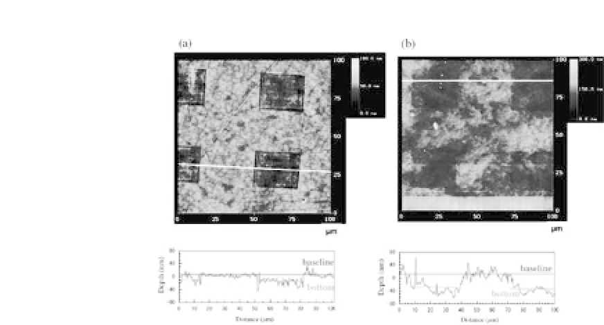
Figure 44.2.
Cross-sectional profiles of the laser-ablated domains on
PIPAAm-1.4 (a) and PIPAAm-2.9 surfaces (b). The laser fluence and the
number of irradiations were 10 mJ/cm2 and 6 times for PIPAAm-1.4 (a)
and 20 mJ/cm2 and 3 times for PIPAAm-2.9 (b), respectively. The baseline
andthebottomwereaveragesofthetextdatafromthesectionprofiles(see
experimental section). The scan size was 100
100 mm
2
. Reprinted with
permission from Ref. 20. Copyright 2004 American Chemical Society.
×
did not contaminate the surfaces of PIPAAm-1.4 and PIPAAm-2.9
with ablated debris. Section profiles showed the depth to be 15.5
±
7.2 nm, indicating the thickness of the grafted PIPAAm layer for
PIPAAm-1.4. Likewise, the averaged thickness of PIPAAm-grafted
layers for PIPAAm-2.9 was 29.3
±
8.4 nm. Considering the amount
and density of the grafted polymer on PIPAAm-1.4 and PIPAAm-
2.9, the measured thicknesses of the grafted polymer layers are
reasonable.
The cell attachment/detachment responses on the PIPAAm-
grafted surfaces are the key factor for noninvasive recovery of
cell sheets for further applications of tissue and organ reconstruc-
tion.Here,weexaminedcellularresponsestothesePIPAAm-grafted
surfaces in terms of cell adhesion and detachment with temper-
ature. Bovine endothelial cells (ECs) adhered and spread on the
PIPAAm-1.4 dishes (Fig. 44.3), whereas they did not on PIPAAm-2.9
(Fig. 44.3). Adhered and proliferated cells on the PIPAAm-1.4 sur-
faces were detached from the surfaces by reducing the temperature
below the PIPAAm's transition temperature for single-cell and/or
monolayer-cellsheets, depending on the cell density of the surface.


Search WWH ::

Custom Search