Biomedical Engineering Reference
In-Depth Information
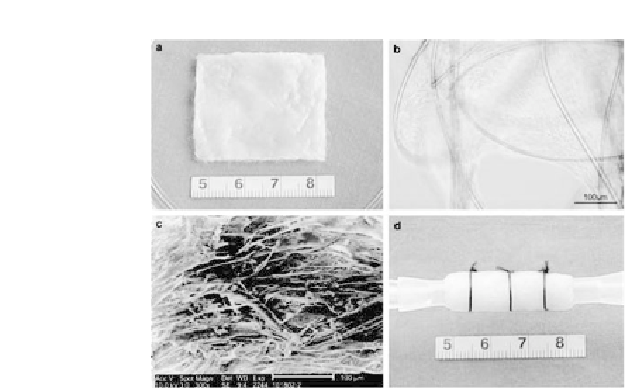
Figure 34.18.
Cell-scaffold constructs cultured in a dish. (a) SMCs were
seeded onto an unwoven PGA fiber mesh and cultured for 5 days in a dish.
(b) Microscopic observation shows SMC growth on the PGA mesh at day 5.
(c) Scanning electron microscopic view of ECM production by SMCs on PGA
fibers at day 5. (d) The cell-PGA sheet was wrapped around a silicone tube
intheculturechamberofavesselreactor,securedbybiodegradablesutures.
(Reprinted by permission from Ref. 20).
dishes. Thereafter, the cell-scaffold constructs were kept in an
incubator for four hours to allow for the complete adhesion of the
cellstothePGAfibers.DMEMwith10%FBSwasthenaddedtocover
the constructs. The cell-PGA sheets were incubated for another five
days in the culture dishes.
Afterward, the SMC-PGA sheets were wrapped around the sili-
cone tubes in the culture chamber of the vessel reactor and fur-
ther secured by biodegradable sutures (Ethilon, Ethicon, Inc., USA)
(Fig. 34.18). The loaded chamber was then filled with DMEM
containing10%FBStocoverthecell-scaffoldconstruct,followedby
connectingtheculturechambertothewholereactor.Apulsatileflow
of sterile PBS was applied through the silicone tubes at a frequency
of75beats/min.Theflowratewasgraduallyincreasedandadjusted
(between 70-80 mL/min) to reach a radial distension about 5% of
the original diameter of the constructs. The culture was maintained


Search WWH ::

Custom Search