Biomedical Engineering Reference
In-Depth Information
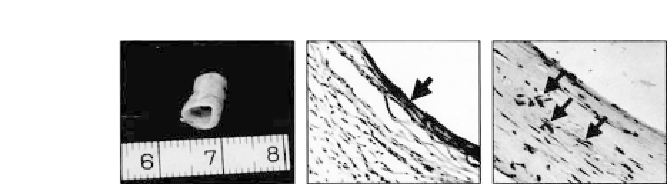
Figure 34.17.
Grossviewofengineeredvesselat6weeks(
left
).Immuno-
histochemistry shows factor VIII-positive cells at the luminal surface (
mid-
dle
)andSM
α
-actin-positive cells in the vessel wall (
right
). Arrows indicate
SM
α
-actin-positive cells. (Reprintedby permission from Ref. 8).
was formed in both groups at 2 weeks but disappeared in the con-
trol group at 6 weeks. Histologically, the implanted PGA fibers were
mostly degraded at 6 weeks (Fig. 34.17). The neovascular structure
formed in the experimental group contained the endothelium on its
luminalsurfaceandatissuelayersimilartothemiddlelayerofaves-
sel. Immunohistochemical staining demonstrated that the endothe-
lium lined at the luminal surface was positive for factor-VIII and
the middle layer contained cells that stained positive for SM
α
-actin
(Fig. 34.17). However, the neovascular tissue harvested at 11 weeks
became atrophic compared with the tissue of 6 weeks, although
trichrome staining showed that more SM fibers were formed in the
11-week tissue than in the 6-week tissue. This phenomenon sug-
gests that mechanical stimulation might be an essential element for
vessel engineering.
To enhance the mechanical property of engineered vessel wall
tissue, an
in vitro
approach was employed using a bioreactor
system.
20
To prepare a cell-scaffold construct, unwoven PGA fibers
(Albany International Research Company, Albany, NY), 15
μ
min
diameter and 30 mg in weight, were made into a 40
×
30
×
2mm
mesh. The scaffold was soaked in 75% ethanol for one hour and
washed three times with PBS, followed by incubating with DMEM
containing 10% FBS for 10 minutes. The medium was removed
afterward,andthescaffoldwasair-driedfor30minutesunderultra-
violet light before use. Canine SMCs were collected, resuspended in
the culture medium at a density of 6
10
7
cells were then evenly seeded onto each PGA mesh in tissue culture
10
7
cells/mL, and 3
×
×


Search WWH ::

Custom Search