Biomedical Engineering Reference
In-Depth Information
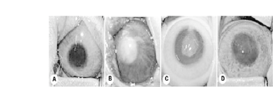
Figure 34.13.
Gross observation shows that engineered tissue gradually
becomestransparentoveran8-weekperiod.(a)Preoperation,(b)postoper-
ation,(c)4weekspostoperation,and(d)8weekspostoperation.(Reprinted
by permission from Ref. 18).
After
in vivo
implantationforeightweeks,theconstructthatwas
opaque at the time of transplantation became a nearly transparent
stroma at eight weeks posttransplantation (Fig. 34.13). The newly
formedstromahasalsobeenprovenasanengineeredstromabythe
presence of green fluorescence protein (GFP)-labeled stromal cells.
Histology of the engineered corneal stroma that was nearly trans-
parent also showed a structure similar to that of the native counter-
part (Fig. 34.14).
18
Inanothersimilarstudy,dermalfibroblastswereusedtoexplore
the possibility of replacing stromal fibroblasts for corneal stroma
engineering.
19
Again PGA fibers were used as the scaffold as sim-
ilarly prepared.
18
Dermal fibroblasts were harvested from new-
born rabbits, seeded onto biodegradable, unwoven PGA fibers, cul-
tured
in vitro
for one week, and then implanted into adult rab-
bit corneas. After eight weeks of implantation, nearly transparent
corneal stroma was formed, with a histological structure similar
to that of its native counterpart (Fig. 34.15). The existence of cells
that had been retrovirally labeled with GFP demonstrated the sur-
vival of implanted cells. In addition, all GFP-positive cells that sur-
vived expressed keratocan, a specific marker for corneal stromal
cells, and formed fine collagen fibrils with a highly organized pat-
tern similar to that of native stroma (Fig. 34.16). Interestingly, the
condrocyte-seeded PGA scaffold formed opaque cartilage instead of
transparent corneal stroma (Fig. 34.15). The results demonstrated
thatneonataldermalfibroblastscouldswitchtheirphenotypeinthe


Search WWH ::

Custom Search