Biomedical Engineering Reference
In-Depth Information
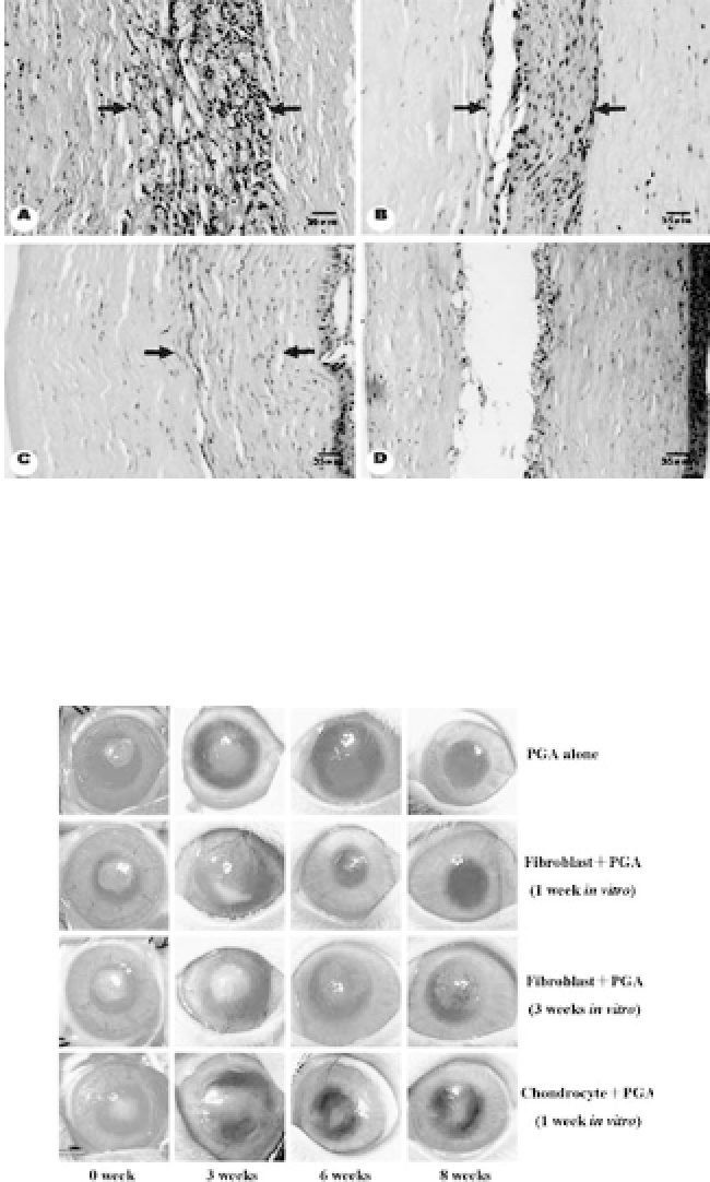
Figure 34.14.
Sectional views of H&E-stained stroma. Implanted corneal
stromal cell-PGA construct regions are indicated by arrows in each exper-
imental group (a, 4 weeks; b, 6 weeks; c, 8 weeks postoperation), and
implanted PGA-alone region in the control group is also shown (d, 8 weeks
postoperation). Original magnification: (a-d)
×
200. Scale bars: 35
μ
m.
Figure 34.15.
Grossviewofthegraftsineachgroupat0,3,6,and8weeks
postimplantation. At 8 weeks, the corneas become transparent in the PGA-
alone group andin thegroup withimplantationof fibroblast-PGAconstruct
precultured
in vitro
for 1 week but not in the chondrocyte-PGA group and
the group implantated with precultured fibroblast-PGA
in vitro
for 3 weeks.
(Reprinted by permission from Ref. 19). See also Color Insert.


Search WWH ::

Custom Search