Biomedical Engineering Reference
In-Depth Information
evaporate freely throughout the vacuum chamber. The atoms and molecules in
the vapor flow then condense onto the substrate surface located at the top of the
chamber, creating a coating that masks the original surface. It was found that
compared with conventional nano-smooth titanium and nano-featured titanium,
osteoblasts adhered and aligned on such nanostructured metal features. Such
observations confirmed the hypothesis that nano-featured titanium can improve
early osteoblast functions (morphology and adhesion), promising for promoting
longer-term functions (such as calcium deposition), criteria necessary to
improve orthopedic implant efficacy [35±37].
Another promising technique that has recently emerged to create nano-
features on titanium is anodization. Anodization is an electrochemical method
also known as anodic oxidation and is widely used as a surface modification
technique for metals to produce protective oxidative layers [38]. Such anodiza-
tion processes usually operate in an acidic solution. Anodization will result in
the formation of an oxide layer on the anode surface. At the same time, the acid
will etch the oxidative layer to cause the initial formation of pores at the weak
points on the titanium surface due to localized dissolution. Then, voids will form
in these inter-pore regions by field-enhanced oxidation/dissolution [39±42].
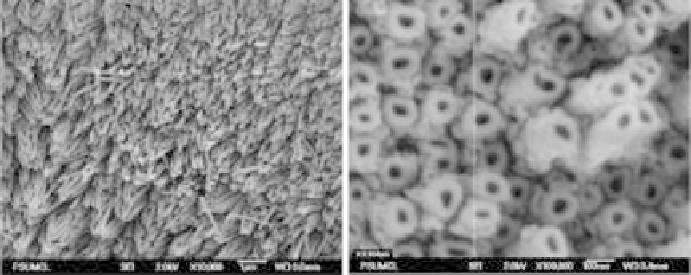
After the anodization of titanium, self-ordered nanotubular surface morpholo-
gies may be observed, as shown in Fig. 9.6.
An in vitro study provided evidence of enhanced calcium deposition by
osteoblasts cultured on anodized titanium with nanotube-like structures
compared with unanodized titanium and titanium anodized to process nano-
particulate structures [43, 44]. A number of in vivo studies have also demon-
strated the promising future of anodized titanium for orthopedic applications.
For example, anodized titanium screws were inserted into rabbit tibia and
allowed to penetrate one cortical layer. After 6 weeks, histology stains showed
bone tissue formation on the nanostructured titanium implant surfaces that had
an oxide thickness of more than 600 nm [45, 46]. Results indicated that the
9.6 SEM images of anodized titanium foil samples. (Adapted from Ruan et al.
[39].)