Biomedical Engineering Reference
In-Depth Information
Forward
Warping
1.16
4
0.90
1
3
2
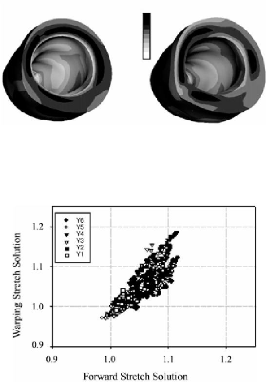
Figure 12.13:
Fiber stretch distribution for the forward (left) and warping
(right) analyses. The locations for the sensitivity analysis are shown on the
forward model as numbers 1-4. Locations 5-8 are at the same locations as 1-4
but at the mid-ventricle level.
Figure 12.14:
Comparison of warping and forward nodal fiber stretch for each
image slice. Y7 corresponds to the slice at the base of LV and Y1 is near the apex
of the LV.
material model. The analysis was repeated and the results compared with the
forward model results.
The forward and Warping sensitivity study results were compared at eight
locations (Fig. 12.3). These results show excellent agreement (Table 3.1) for
all cases indicating hyperelastic Warping is relatively insensitive to changes to
material model and material parameters. These results indicate that accurate
predictions can be determined even when material model and parameters are
not known. This is consistent with our previous results of Warping analyses of
intravascular ultrasound images [22].