Biomedical Engineering Reference
In-Depth Information
12.3.4.1
Validation of Warping for Tracking Left Ventricular
Deformation using Volumetric MRI
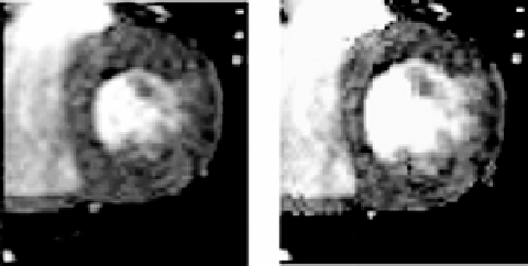
To validate the use of Warping for predicting LV strains from sets of volumetric
cine-MRI images, a pair of 3-D cine MRI image datasets representing two states of
the left ventricle during the cardiac cycle was required. Further, the deformation
map between the states represented in the images had to be known to provide a
gold standard for comparisons. This was achieved by first acquiring a gated 3-D
cine-MRI dataset of a normal volunteer's heart during early diastole on a 1.5T
Siemens scanner (256
×
256 image matrix, 378 mm FOV, 10 mm slice thickness,
10 slices). This volumetric MRI dataset was designated as the
template
image
(Fig. 12.11, left). The endocardial and epicardial surfaces of the LV were hand
segmented. An FE model of the left ventricular (LV) image space was created
based on these segmentations (Fig. 12.12, left panel). The myocardium was
represented as a transversely isotropic material with the fiber angle varying
linearly from
−
90
◦
at the epicardial surface, through 0
◦
at the Mid-wall, to 90
◦
at the endocardial surface [91]. The material coefficients were determined by
least squares fit of the transversely isotropic hyperelastic constitutive model
described in Weiss et al. [30] described above in the intravascular ultrasound
section, to the biaxial stress/strain values presented in the work of Humphrey
et al. [31, 32].
An internal pressure load representing end-diastole was applied to the lu-
men and a standard “forward” nonlinear FE analysis was performed using the
Figure 12.11:
Mid-ventricular slices of the template (left) and the target (right)
image datasets used in the validation analyses. Left image was obtained from
direct MR volumetric image acquisition, while right image was created by de-
forming left image using results of forward FE analysis (see text).