Biomedical Engineering Reference
In-Depth Information
involve the use of neat PEEK for both cervical and
lumbar spinal cages
[37
e
47]
. A tapered PEEK cage
for the lumbar spine is shown in
Fig. 13.11
. Because
the use of neat PEEK for spine is a relatively recent
development, the published literature is generally
limited to in vitro biomechanical studies
[43
e
45]
,or
short-term outcomes in animal studies or human
clinical trials
[37
e
41,46,47]
. Recent studies with
PEEK cages have looked to improve or accelerate
fusion performance by combining the devices with
the use of hydroxylapatite
[41]
, 40%
b
-tricalcium
phosphate/60% hydroxylapatite
[38]
, or rhBMP-2 on
a collagen sponge
[40]
.
A detailed animal study investigating PEEK
interbody cages has recently been reported by Toth
et al.
[40]
. Researchers studied the influence of
rhBMP-2 on a collagen sponge (InFuse
: Medtronic
Spinal and Biologics, Memphis, TN) on fusion with
a PEEK-threaded cage in an ovine model. An
autograft group served as a control (
Fig. 13.12
).
After 6 months, the biomechanical behavior of
the treated level was measured. The cage group
treated with InFuse
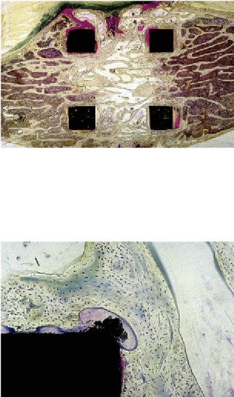
Figure 13.9
Histology confirmed the radiographic find-
ings of fusion in the cages of the Spanish goat study.
Extensive growth of trabecular bone was observed
through the cages. Image courtesy of Bill Christianson,
DePuy Spine.
was found to be significantly
stiffer in flexion and left lateral bending than the
control group treated using a PEEK cage filled with
autografts. Histology and microradiographs were
used to characterize the extent of fusion (
Fig. 13.13
).
All the PEEK cages with InFuse
were diagnosed
with fusion at 6 months radiographically and histo-
logically. No evidence of wear debris or damage to
the device was noted. Only mild inflammatory
response was noted during histological evaluation of
tissue adjacent to the PEEK implants, which the
authors judged to represent “excellent” biocompati-
bility for the material.
Intermediate-term clinical reports on the use of
unfilled, neat PEEK cages are reported in the spine
literature
[48]
. A retrospective study of neat PEEK
cages used for circumferential lumbar fusion was
reported by Rousseau et al.
[48]
, who evaluated 57
consecutive patients after a mean follow-up of 5.7
years (range: 4
e
8 years). They observed fusion in
56 cases, but they were unable to maintain the desired
curvature of the spine in 10 patients. They attributed
the loss of spinal curvature correction to the order of
implantation of the cages, which occurred after the
spine was locked in place using a rigid posterior
instrumentation system. The authors concluded that
“lumbar circumferential arthrodesis using PEEK
cages
.
provided good clinical results and fusion
rate.”
Figure 13.10
Histology confirmed the absence of an
inflammatory reaction to the CFR-PAEK in the Spanish
goat model. The black particles at the corner of the
cage strut may be polishing artifact, rather than wear
debris. No foreign body reaction is observed. Image
courtesy of Bill Christianson, DePuy Spine.
Brantigan cages were originally machined from
CFR-PAEK plates. Both 30% PEKEKK and 70%
continuous carbon fibers, as well as 70% PEKEKK
with 30% carbon fiber, were originally used
[36]
.
Today, these cages are fabricated more efficiently by
injection molding CFR-PEEK OPTIMA (70% PEEK
with 30% chopped carbon fiber)
[36]
.
13.4 Threaded PEEK Lumbar
Fusion Cages
Although many articles describe the use of CFR-
PEEK for spine implants, many recent studies also