Biomedical Engineering Reference
In-Depth Information
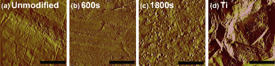
Figure 10.5
AFM images of the injection-molded PEEK: (a) unmodified, relatively smooth surface; (b) 600 s modified,
showing little change in the surface roughness; (c) 1800 s modified PEEK showing pits created as a result of the longer
treatment time. The change in topography observed is due to the “etch effect” of prolonged exposure to oxygen
plasma; this pitting could potentially provide anchorage points for osteoblast cell adhesion. (d) Micro-rough medical
grade titanium showing a much greater surface roughness than that observed on the PEEK surfaces (10 mm
scale bar).
a period of 26 months, the surface chemistry and
wettability of our oxygen plasma-modified PEEK
surfaces have been evaluated and shown to be stable.
Oxygen plasma treatment is known to affect surface
topography; we therefore evaluated the modified and
unmodified PEEK surfaces by atomic force micros-
copy (AFM).
AFM analysis of the untreated PEEK, presented in
Fig. 10.5
a, showed the residual striations from the
injection mold, where the maximum peak to trough
distance observed was under 300 nm, and the stria-
tions were relatively irregular. Following 600 s of
plasma treatment, as presented in
Fig. 10.5
b, the
surfaces were still quite similar to the surfaces of the
unmodified PEEK, although the peaks of the striations
were slightly less sharp. Minor changes to the surface
roughness were observed on all the surfaces modified
for less than 1200 s (data not shown); however, those
surfaces modified for longer had a very apparent
change in the topography, where the etch effect of the
plasma treatment was obvious, clearly showing the
striations to be less prominent, leading to a more
uniformly rough surface. After 1800 s of exposure to
oxygen plasma, the etching of the surface was very
apparent, shown in
Fig. 10.5
c, where the surface
topography has clearly changed, and the original
striations were hardly visible; these pits were also
confirmed by scanning electron microscopy (SEM).
The surface roughness of all the surfaces showed that
all the PEEK surfaces were relatively smooth in
comparison to standard microrough commercially
pure titanium (Ti; ISO 5832/2; Synthes, CH). The
etching effect of the plasma treatment did not signif-
icantly change the surface roughness of the modified
PEEK surfaces, although the surfaces from the longer
treatment times do have a slightly higher average
roughness. Having confirmed the increase in surface
oxygen, change in topography and increase in surface
energy, the surfaces were evaluated for their cyto-
compatibility with human primary osteoblast-like
(HOB) cells evaluating cellular attachment, prolifer-
ation, morphology, and characteristic behavior over
28 days in vitro culture.
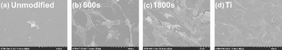
Figure 10.6
shows the HOB
cell adhesion 1 day postplating.
Assessing the cell density and thereby prolifera-
tion on the various sample surfaces allows the effect
Figure 10.6
Scanning electron micrographs showing HOB cell attachment 1 day postplating to the (a) unmodified,
(b) 600 s, (c) 1800 s modified PEEK, and (d) microrough medical grade titanium (Ti). There was much greater cell
adhesion and spreading to the modified PEEK and Ti surfaces (b, c, d) in comparison with unmodified PEEK (a), where
the cells appear to be lifting off.