Biomedical Engineering Reference
In-Depth Information
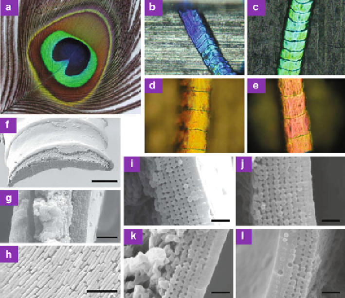
Fig. 8.29
(
a
) Eye pattern of a tail feather of a male green peacock
P. muticus
.(
b
)-(
e
) Optical
microscopic images of
blue
(
b
),
green
(
c
),
yellow
(
d
), and
brown
(
e
)barbules.(
f
)-(
l
) SEM images
of barbules. (
f
) Perspective view of a sectioned yellow barbule. (
g
) Transverse cross-section of
a green barbule. (
h
) Longitudinal cross-section of a green barbule with the surface keratin layer
removed. Melanin rods can be clearly seen. (
i
)-(
l
) Transverse cross-sectional images of
blue
(
i
),
green
(
j
),
yellow
(
k
), and
brown
(
l
) barbules. Scale bars: (
f
)10
m; (
g
)2
m; (
h
)1
m; and (
i
)-(
l
)
500 nm
are air holes. Melanin rods are parallel to the cortex surface, running along the axis
of a barbule. Photonic-crystal structures in all differently colored barbules are quite
similar. In the blue, green, and yellow barbules, the lattice structure is a square
lattice, whereas in the brown barbule it is a rectangular lattice. The only differences
are the lattice constant (rod spacing) and the number of periods (melanin rod layers)
along the direction normal to the cortex surface. The lattice constant for the blue,
green, and yellow barbules is 140, 150, and 165 nm, respectively. In the brown
barbule, the lattice constant is 150 and 185 nm along the directions parallel and
perpendicular to the cortex surface, respectively. The number of periods is 9-12 for
the blue and green barbules, and about 6 for the yellow barbules. The brown barbules
have the least number of periods, about 4. These differences in the lattice constant
and number of periods in differently colored barbules can give rise to diversified
coloration.