Biomedical Engineering Reference
In-Depth Information
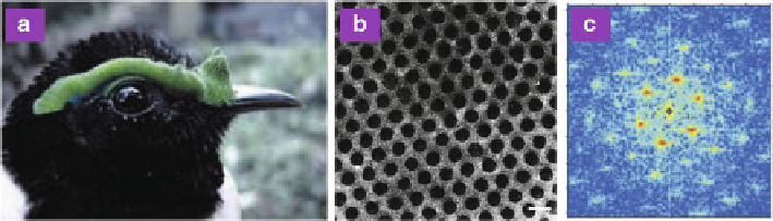
Fig. 8.28
(
a
) Optical image of
P. castanea
.(
b
) TEM image of the nanostructured arrays of the
dermal collagen fibers. (
c
) 2D Fourier power spectrum of the TEM image showing discrete bright
points of spatial frequency. Scale bar: (
b
) 200 nm (Reproduced from [
121
])
Structural blue and green colors occur in the skin of a broad diversity of birds
from many avian orders and families. Structural characterizations with electron mi-
croscopy revealed that the dermal collagen layer contains quasi-ordered or ordered
arrays of parallel collagen fibers [
121
], which are responsible for the skin coloration.
The arrays of collagen fibers in the skin of most birds are quasi-ordered, but in
Philepitta castanea
(Eurylaimidae) the collagen fibers are exceptionally ordered,
arranged in a hexagonal array, as shown in Fig.
8.28
. This ordered arrangement
of the collagen fibers manifests a 2D hexagonal photonic crystal, confirmed by a
hexagonal pattern of spatial frequency peaks in the power spectrum, obtained from
a 2D Fourier analysis.
The most remarkable example of 2D photonic crystals in the biological world
resides in peacock feathers [
122
-
125
]. Over the centuries, humanity has been
impressed by the splendor of the colors of peacocks. The coloration of peacock
feathers had puzzled scientists for a long time. More than 300 years ago, Hook and
Newton studied peacock feathers by optical observations and suggested that their
colors were produced by thin films. The ultimate physical mechanism of the color
production was, however, uncovered only recently [
124
,
125
].
The male peacock tail contains spectacular beauty because of the brilliant,
iridescent, diversified colors, and the intricate, colorful eye patterns. In the eye
pattern of a tail feather of a male green peacock (
Pavo muticus
), blue, green, yellow,
and brown feathers can be found, as shown in Fig.
8.29
. A typical peacock tail
feather possesses a central stem with an array of barbs on each side. Barbs are
colorless. On each side of a barb there is an array of flat barbules. Perceived feather
colors are from barbules. Each barbule has connected round indentations of typically
about 20-30
m. The round indentations have a smoothly curved crescent-like
profile in transverse cross-section. From transverse cross-sectional SEM images, a
barbule consists of a medullary core of about 3
m enclosed by a cortex layer. The
medulla consists of randomly dispersed keratin and melanin. In contrast, the cortex
layer displays a regular structure. The outer surface of the cortex is a thin layer of
keratin. Beneath the surface keratin layer, there is a 2D photonic-crystal structure,
made up of an array of melanin rods connected by keratin. The remaining hollows