Biomedical Engineering Reference
In-Depth Information
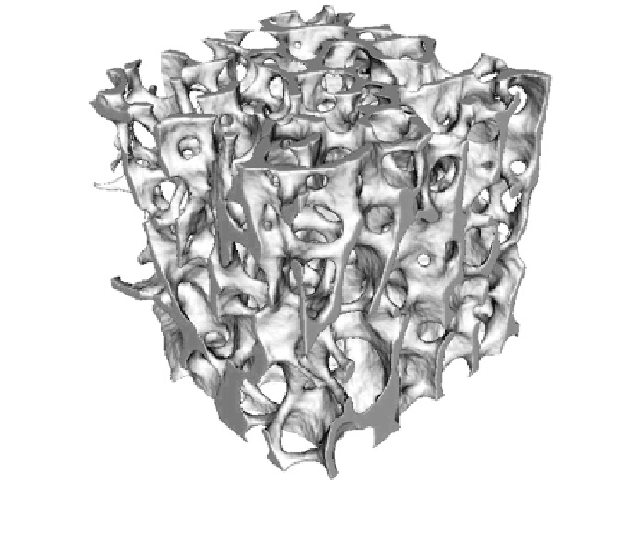
Fig. 5 3D rendering of a bone cube from the L4 vertebral body of 66 years old male with
BV/TV = 9.6%
improving, is problematic and there are limited facilities in which large clinically-
relevant bone samples can be imaged. In addition, synchrotron-derived datasets
present challenges in data handling, where it is not unusual for a single sample to
generate at least 100 GB of data, whereas a dataset from a laboratory-based micro-
CT system will be less than 10 GB in size. These massive synchrotron datasets
require computing resources not commonly available. For non-ionizing radiation
imaging, magnetic resonance (MR) imaging is approaching the spatial resolution
of micro-CT, where in-plane resolution is approaching 100 lm and apparent
resolution below 100 lm with sub-voxel processing techniques [
59
]. However,
compared to micro-CT imaging there are still limitations in the ability to accu-
rately delineate the mineral and non-mineral phases, which have limited the
adoption of this imaging modality for morphometric bone studies [
57
,
59
,
61
,
67
].
One of the challenges of three-dimensional imaging of trabecular bone by
whatever imaging modality is the ability to manipulate the wide range and large
volume of data acquired from imaging, for 3D reconstruction and for morpho-
metric analysis. However, consumer-level computers are able to process desktop
micro-CT datasets reasonably efficiently, in terms of processing time, data gen-
eration and data storage needs.
Search WWH ::

Custom Search