Biomedical Engineering Reference
In-Depth Information
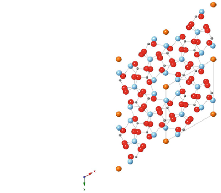
Fig. 1 Chemical diagram of
2
3
unit cell of hydroxyapatite
shown in the 001 plane.
Hydroxyapatite has
hexagonal structure of P63/m
and has a unit cell outlined in
the figure with dimensions of:
a = 9.432 Å, b = 9.432 Å,
and c = 6.881 Å, with
calcium (Ca, gray),
phosphate (P, blue), oxygen
(O, red), and hydroxyl (OH,
orange). (Generated by Jared
Diegmueller [
56
] using
Crystal Maker software)
(DXA) is the most widely applied technique to quantify BMC/BMD changes with
age or osteoporosis, although the BMD measured by DXA represents areal BMD
(aBMD = BMC/projected area). Quantitative computed tomography (QCT)
allows determination of a volumetric BMD (vBMD), a volumetric measure of
bone mineralization. vBMD is a measure inclusive of porosity (i.e. haversian
canals, vascular channels). On the other hand, degree of mineralization (DOM) or
specific mineralization at the solid tissue level, is measured by methods which
resolve bone below microporosity, such as microradiography, ashing, back scat-
tered electron microscopy or spectroscopy (FTIR, Raman). Therefore, the quantity
of bone mineral and how mineralized the bone matrix are, represent two separate
issues which require multiple methods to be addressed specifically.
During osteogenesis, the initial stages, unmineralized osteoid is deposited. The
osteoid is composed of type I collagen, non-collagenous proteins, proteoglycans
and water. Mineralization occurs with some delay within the continuum of the
osteoid (Fig.
2
), presumably by displacing water. This theory is indirectly sup-
ported by the study of Mueller et al. study which reported increased degree of
mineralization in elderly with decreased water content [
113
].
The osteoid turns into fully mineralized bone during two phases. During
primary mineralization, mineral crystals grow and agglomerate to form bigger
Search WWH ::

Custom Search