Biomedical Engineering Reference
In-Depth Information
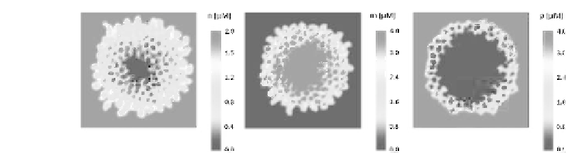
FIGURE 8.15: (See color insert.) Final patterns (i.e., at t = 15 days) of the
microscopic variables included in the model, taken in a representative section
of the domain. From left to right panel: concentration of growth factors (n),
tumor proteolytic enzymes (m), and matrix proteins (p). It is straightforward
to notice that cells in the center of the spheroid have a negligible amount of
chemicals and therefore undergo the necrotic transition. The more external
individuals have instead an abundant supply of vital nutrients, which increase
their metabolism. Indeed, the extracellular environment is completely modi-
fied by the tumor, as the matrix components are degraded by the localized
activity of malignant MMPs.
matrix proteins. In particular, it can be clearly observed that the host tissue
is significantly modified, as the matrix components have been dramatically
degraded by the activity of tumor proteolytic enzymes. Indeed, steep protein
gradients have formed, which eventually drive, via haptotactic mechanisms, a
further invasion of the single, aggressive individuals.
The evolution of the malignant mass captured in the model reproduces
the first stages of the growth of several tumors grown as spheroids in spinner
cultures, such as ovarian [53, 363] or breast [155] carcinomas. Moreover, the
model results are consistent with the development of avascular gliomas , both
embedded in vitro in collagenous gels [208, 376] and implanted in vivo in
mice [2, 69]. It is useful to underline that such a qualitative agreement occurs
even without an exact bookkeeping of diffusing growth factors and a detailed
inclusion of the cell cycle. Indeed, a similar growth of primary solid tumors
have been also predicted by different types of theoretical models, see again
the reviews [15, 58, 74, 321] and the comprehensive topics [240, 316].
As briefly sketched in the previous section, the values of the parameters
Js have a clear biological relevance, as they describe the relative preference
of tumor cells to be in contact with other cells or with matrix components.
At a molecular level, they are in fact a measure of the expression and the
engagement of the different types of cell adhesion molecules, cadherins and
integrins, respectively. As the variation of the Js may be expected to have a
substantial impact on the overall development of the tumor, we analyze the
model outcomes obtained by varying the cell{cell adhesive strength, dened
by J
C;C
. As reproduced in Figure 8.16, at low values of J
C;C
, which mean


Search WWH ::

Custom Search