Biomedical Engineering Reference
In-Depth Information
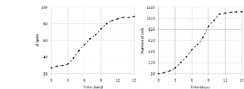
FIGURE 8.14: Regression of the time-evolution of both the invasive radius
d (left panel) and the number of cells (right panel) for the tumor spheroid,
developing under the standard parameter settings, as depicted in Figure 8.13.
intercellular adhesive forces and the physiological regulation of the contact-
inhibition of movement, inducing a sort of mesenchymal transition. These shed
individuals, which are dicult to be clinically detected, have the greatest po-
tential to invade the host and further metastasize. As seen in the previous sec-
tions, they display in fact an evident ability to wonder in the close proximity
and to spread in the surrounding tissue, mainly driven by haptotactic mech-
anisms via local ECM gradients created by the activity of their proteolytic
enzymes. Evading destruction by the immune system, they may subsequently
establish secondary colonies with devastating consequences for the wellbeing
of the patient, as the likelihood of success of therapeutic interventions strongly
decreases [162, 324].
A specific description of the cell phenotypic differences within the spheroid
is obtained by considering the distribution of the intracellular levels of growth
factors, given in Figure 8.15 (left panel). Cells in the more external ring of the
tumor have an abundant supply of chemicals and, as seen, can freely proliferate
and eventually shed in the extracellular environment, as a consequence of
their increased metabolism and motility. As the distance between the spheroid
border increases, the local availability of growth factors progressively falls.
Indeed, a sort of equilibrium point is reached at which cells have an internal
amount of vital factors too low to undergo mitosis, but suciently high to
survive. These individuals are therefore in a so-called quiescent state. Finally,
in the core of the mass, the supply of chemicals is negligible, so that inner
cells, once consuming their basal level of growth factors, are unable to stay
alive. They therefore die due to nutrient deprivation forming, as seen, a region
of necrotic debris, which loose volume and are eventually removed. Summing
up, malignant individuals within a solid, avascular mass display three different
phenotypic state: proliferating, quiescent, and necrotic.
Figure 8.15 represents also the final distribution of tumor MMPs and of


Search WWH ::

Custom Search