Biomedical Engineering Reference
In-Depth Information
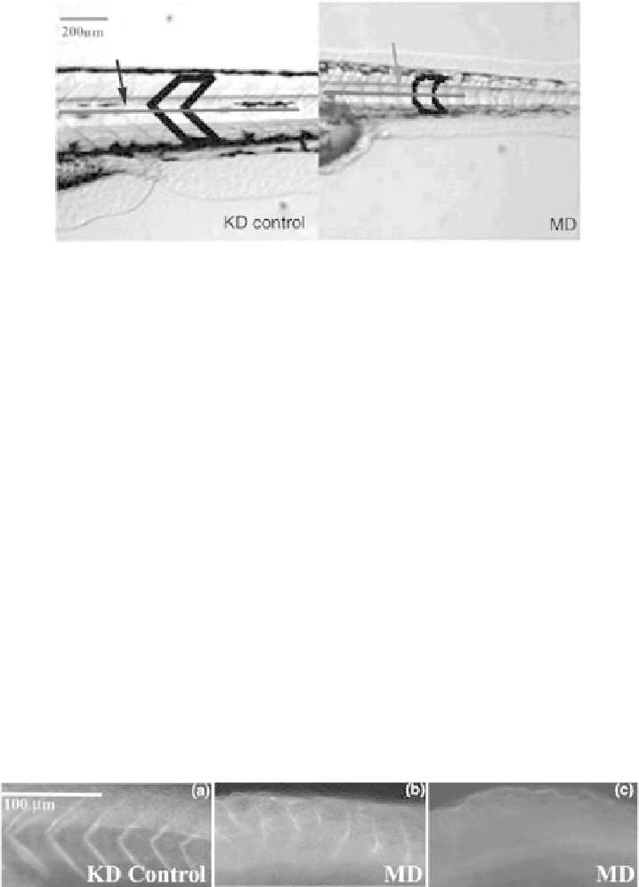
Figure 18.2
Myotome structure. Three dpf zebrafish tails are shown (3.2 magnification).
In KD control zebrafish, myotomes in the tail exhibited the characteristic chevron, V-shape (black arrow;
thick black outline), whereasMDzebrafish exhibited defectivemyotomes (red arrow; thick black outline).
Blue scale bar indicates 200 mm. Blue lines span myotomes used for quantitation in Fig. 18.4.
zebrafish decreased after 5dpf, we decided to focus on MD phenotype development
from 1 to 5dpf (Fig. 18.1).
Uninjected zebrafish and KD controls exhibited normal body morphology,
whereas MD zebrafish exhibited small heads (green arrows), small eyes (yellow
arrows), pericardial edema (blue arrows), truncated bodies (red arrows), and short or
bent tails (black arrows, Fig. 18.1). At 2, 3, and 4dpf stages, difference in overall body
and tail length, from the anal pore to the tip of the tail (Fig. 18.2), was significant.
18.3.2 Confirmation of Dystroglycan Knockdown
by Whole Mount Immunostaining
To confirm that MD phenotypes resulted from dystroglycan knockdown and not from
nonspecific siRNA effects, we performedwholemount immunostaining using an anti-
human dystroglycan antibody shown to cross-react in zebrafish (Parsons et al., 2002).
Staining pattern showed low (Fig. 18.3b) or loss of (Fig. 18.3c) dystroglycan
expression and abnormal sarcolemma.
Figure 18.3
Whole mount dystroglycan immunostaining. Twenty hpf zebrafish were stained with a
dystroglycan-specific antibody. (a) KD control zebrafish exhibited dystroglycan staining in the
myoseptum of normal myotomes, whereas MD zebrafish exhibited either (b) weak dystroglycan
staining in irregular shaped myotomes or (c) absence of dystroglycan staining. These results confirmed
KD of dystroglycan expression by siRNA. Fluorescence images were captured at 4
magnification;
anterior, left. White scale bar represents 100 mm.
Search WWH ::

Custom Search