Biomedical Engineering Reference
In-Depth Information
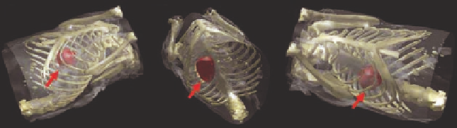
FIGURE12.9: Multimodality imaging. The fluorescent reconstructions of
Figure 12.8 rendered simultaneously with X-ray CT images. The tumor is
indicated by an arrow. (From [31].)
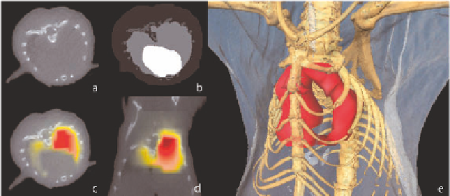
FIGURE12.11: Reconstruction based on data from combined FMT-XCT
setup. (a) X-ray slice. (b) Segmentation of X-ray data in lungs, heart, bone
and remaining tissue. (c) Reconstruction of fluorescent biodistribution in lung,
transversal slice and (d) sagittal slice. (e) 3D-hybrid visualization. (From [22].)