Biomedical Engineering Reference
In-Depth Information
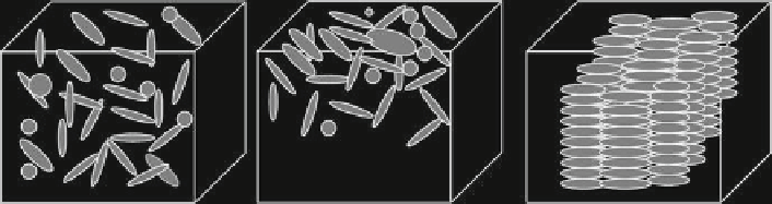
Fig. 4
Schematic showing the lack of control provided by current scaffolding techniques and the
type of control needed for true tissue engineering. (
Left
) Cells are mixed into the matrix or (
Center
)
cells are seeded on top of the matrix. Neither of these approaches provides control over cellular
organization. (
Right
) Cells show a precise, controlled 3D organization in the matrix. This is not
possible with current scaffolding techniques. Cells are represented by
gray ovals
creating unorganized groups of cells to a 3D-engineered tissue with a high degree of
organization at the level of individual cells (Fig.
4
). A scaffold that could induce
proper cellular organization in the x, y and z directions would allow for the construc-
tion of artifi cial tissues that recapitulate the properties of the natural counterpart.
It has been shown that arrays of microgrooves on culture surfaces can organize
monolayers of cells in the direction of the pattern. This was accomplished by taking
advantage of the phenomena of contact guidance that has been shown to provide con-
trol over the organization of cells in two-dimensional cultures (Dunn and Heath
1976
) .
Contact guidance has been demonstrated to have an infl uence with many different cell
types including fi broblasts (den Braber et al.
1998
), cardiac myocytes (Deutsch et al.
2000
), smooth muscle cells (Sarkar et al.
2005
), neurons (Degenaar et al.
2001
) and
bone marrow cells (Matsuzaka et al.
2003
). All of these studies have shown that sur-
face topography plays an important role in cellular organization. The texture of the
surface has also been shown to have signifi cant infl uence on fi broblast adhesion and
proliferation (van Kooten et al.
1998
). These observations led us to design an extension
of this system that would provide similar control over organization in three dimen-
sions. A three-dimensional artifi cial tissue system was created that has organization at
the cellular level similar to that which would be found in a native in vivo system.
The design of the scaffold is a microfabricated 3D array of deep, parallel chan-
nels that is integrated into the interior of a collagen matrix. The ordered pattern of
polydimethylsiloxane (PDMS) acts as a skeleton to guide the cells growing in the
collagen matrix (Fig.
5
). The channels are created in PDMS using standard photoli-
thography and soft lithography techniques. A collagen gel seeded with fi broblasts is
then molded around the PDMS skeleton and the entire construct is cultured under
standard cell culture conditions. By using the natural collagen matrix to suspend the
cells, the cells are provided with an ECM environment that is similar to what would
be found in the body while the PDMS internal skeleton provides a physical barrier
that the cells respond to by organizing in relation to the channels.
Cellular parameters varied signifi cantly between control gels and scaffolds. Of
most interest was the orientation of the cells in relation to the scaffold. Control gels,