Biomedical Engineering Reference
In-Depth Information
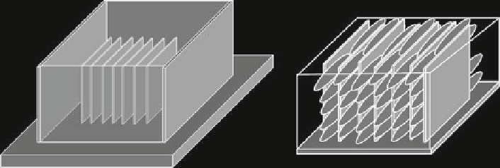
Fig. 5
Schematic showing desired effects of using an internal scaffold to control three-dimen-
sional cellular organization in a matrix. (
Left
) The PDMS scaffold design. (
Right
) The pattern of
cellular organization created using this design
which contain no internal skeleton to provide contact guidance, allowed cells to
grow randomly throughout the gel. No underlying organizational pattern is observed.
With the addition of an internal scaffold to control cell migration, the organization
of the cells becomes strikingly clear. The cells were shown to be highly aligned with
the pattern of the scaffold at every level in which they appeared within the three-
dimensional construct as seen by confocal fl uorescence microscopy (Fig.
6b
). While
this system was used as a model for making a composite scaffold that can guide cell
growth, it does have physiological relevance to tissue systems such as tendon and
muscle which have similar cellular organization. In addition, cell morphology was
found to differ in the scaffolds, where cells were longer and thinner than those
grown in control gels. This is presumed to be because of their interaction with the
PDMS skeleton. It is still unclear how the cells are interacting with the PDMS walls
of the scaffold. They may be treating it as a surface and growing along it as if they
were on a fl at, 2D culture turned on its side. The reduction in the observed minor
axis and the increased spread in the major axis may be indicative of the cell fl at-
tening and spreading on the wall. In this case, this skeleton could provide further
surface area for nanoscale modifi cation for the cells to interact with.
As it is, the fusion of a scaffold with a precisely defi ned architecture with a
matrix that can support cell growth led to an engineered construct that takes on a
three-dimensional organization not possible with standard 3D culture techniques.
Further work needs to be done in order to assess how differing scaffold and matrix
parameters affect the cells. One area of particular interest is the use of biodegrad-
able polymers to replace the PDMS of the scaffold. PDMS serves as a model sys-
tem, but once the cells proliferate and organize, it would be desirable to remove the
internal scaffold. This could be accomplished with the use of a biodegradable poly-
mer designed to degrade at some point after the cells organize. Also, the incorpora-
tion of pores into the PDMS skeleton could allow better diffusion of nutrients to
cells. This should be done without disrupting the architecture of the pattern itself as
to not lose the control over organization provided. Methods for accomplishing this
are addressed in the next segment.