Biomedical Engineering Reference
In-Depth Information
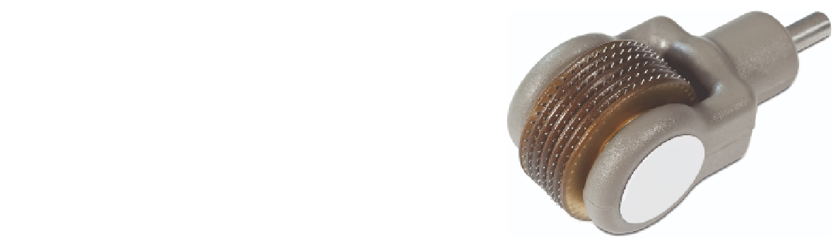
Figure 1.28
Tip of diode-radiofrequency device (metal rails (
arrows
) are the
electrodes in bipolar confi guration; Comet, Syneron, Canada).
Figure 1.29
Radio frequency fractional handpiece; the small electrodes create
plasma on skin as device rolls over surface.
a brief overview of applications
Psoriasis
In psoriasis, one can target the microvasculature with vascular-
specifi c lasers or, alternatively, vaporize plaques with resurfacing
lasers. Owing to the size of the vessels, the PDL with its shorter
pulse is the most logical choice among vascular lasers (83-85).
Laser resurfacing has also been used to remove lesions (86).
The excimer laser has been used. Here photochemistry is the
primary mechanism (87). PDT confi gurations have also been
applied with varying degrees of plaque clearing (88-90).
the tissue heating is determined by variations in electrode type,
power, and cooling times (75). If one uses “rail” metal-type
electrodes placed next to a fl ashlamp crystal, EM fi eld theory
predicts that there will be a hot spot near the edge of the elec-
trode. These hot spots can be reduced by electrically coupling
the energy into the skin (e.g., ensuring that the dry stratum
corneum, with high intrinsic impedance, is wetted with an
electrolyte solution).
One device (Aurora SR, Syneron, Richmond Hill, Ontario,
Canada) combines RF energy and a fl ashlamp. The near-
simultaneous application of electrical and optical fi elds is
proposed to optimize effi cacy and safety. In this confi gura-
tion, the local optical energy (fl uence) increases the discrete
chromophore temperature (i.e., hair, vessel). This localized
heating reduces impedance (skin is treated as an electrolyte
with decreasing impedance as a function of increasing tem-
perature) and therefore in higher localized current densities.
Thus, there is “synergy” between the optical and electrical
parts of the device (75). A purported advantage of the treat-
ment is that lower optical energies can be used to selectively
heat subsurface targets than if a light source were used alone
(thus enhancing epidermal preservation). There is some evi-
dence that white hairs could be reduced with this technology.
The working theory is that although there is little melanin in
white hairs, there is higher current density around the follicle
(as the current navigates around the high-resistance shaft)
(75,77,78) The current density in the tissue adjacent to the
hair shaft is roughly twice the current density and other parts
of the skin; this is because the electrical current streams
around the hair shaft concentrating in a layer just around the
hair. Although certainly promising in principle and based on
sound scientifi c principles, no study has clearly shown at the
time of this writing that the “synergy,” at least in its present
confi gurations, is clinically relevant. That is, for example, no
peer-reviewed study has shown that hair removal without RF
is more effective than that with RF (with all other parameters
held constant). Many studies that may or may not support the
role of RF energy and optical energy as good dance partners
are pending.
More recently fractional RF devices have been introduced
for skin rejuvenation. Both bipolar and monopolar designs
have been applied to create microwounds at and just below the
skin surface (Fig. 1.29) (79-82).
Hypopigmentation
The excimer laser and UV lamp sources have been used to
restore pigment through photochemical pathways (48,91).
Postinflammatory Hyperpigmentation
This is typically very resistant to laser therapy. Some excep-
tions are Q-switched laser therapy for long-standing hyperpig-
mented areas on the extremities. Also, the PDL, in treating
certain hyperpigmented hypertrophic scars, can reduce both
the pigmentation and vascular aspects of the scar. Finally,
long-pulsed KTP lasers and IPLs can sometimes reduce postin-
fl ammatory hyperpigmentation.
Wrinkle and Scar Reduction
They are typically achieved via ablative mechanisms. The CO
2
and Er:YAG lasers are ideally suited for LSR. The CO
2
laser func-
tions more as a heating tool with typical parameters (5-10 J/cm
2
in pulsed mode), whereas the Er:YAG laser acts more as a purely
ablative tool. A summary of the mechanisms follows. In brief,
when energy deposition occurs rapidly, water does not vaporize
at 100°C because the pressure is higher than 1 atm. Energy is
deposited isovolumetrically and the temperature may reach
300°C with pressures up to 1000 atm (19). These high-pressure
gradients can assist tissue removal because, depending on the
mechanical properties of the tissue, the explosive removal pro-
cess can be more energetically effi cient than the 2500 J/cm
3
latent heat of vaporization for water. For example, the heat of
ablation for epidermis is small, and portions of the friable cel-
lular epidermis can be observed in the plume. Thus not all of
the tissue is actually vaporized but rather forcibly ejected from
the surface. For dermis, about 4.3 kJ/cm
3
are required for abla-
tion by many CO
2
lasers—almost twice that needed for water