Biology Reference
In-Depth Information
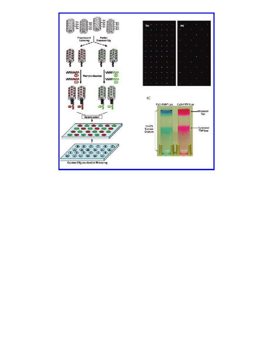
Figure 7.17
TMV microarrays. Left panel: hybridization-based programming of
fluorescently labeled and partially disassembled TMV nanotemplates for assembly
onto multiple addresses on DNA oligonucleotide microarray platforms. Right panel:
patterned assembly of fluorescently labeled and programmed TMV nanotemplates
onto oligonucleotide microarrays via hybridization of Cy5-TMV1cys-D and Cy3-
TMV1cys-H simultaneously. Cy5 and Cy3 are fluorescent dyes, the TMV particles
used were Cys-added mutants referred to as TMV1cys, and D and H denote different
oligonucleotide specificities. (a) Alternating pattern of sequences D and H. (b)
Alternating pattern of sequences G and H. (c) Sucrose gradient centrifugation
of Cy5- and Cy3-labeled TMV nanotemplates. Unreacted dyes remain at the top,
whereas labeled TMVs appear in the middle as distinctive bands. Reproduced with
permission from Yi, H., Nisar, S., Lee, S. Y., Powers, M. A., Bentley, W. E., Payne, G. F.,
Ghodssi, R., Rubloff, G. W., Harris, M. T., and Culver, J. N. (2005) Patterned assembly of
genetically modified viral nanotemplates via nucleic acid hybridization,
Nano Lett.
,
5
(10), 1931-1936.
TMV has been specifically immobilized on surfaces via nucleic acid
hybridization (Tan
., 2005, 2007). The polar nature of
TMV was utilized. The helical encapsulation of the RNA molecule results in
sequence-definable 5
et al
., 2008; Yi
et al
ends. A mild disassembly protocol was used
to partially disassemble the protein coat and expose the RNA at the 5
′
and 3
′
′
end.
Search WWH ::

Custom Search