Biology Reference
In-Depth Information
A
N1234 C
B
N 1234C
Lysate/Anti-tag
C
N
1
2
3
4
C
Sup/Anti-tag
Lysate/CBB stain
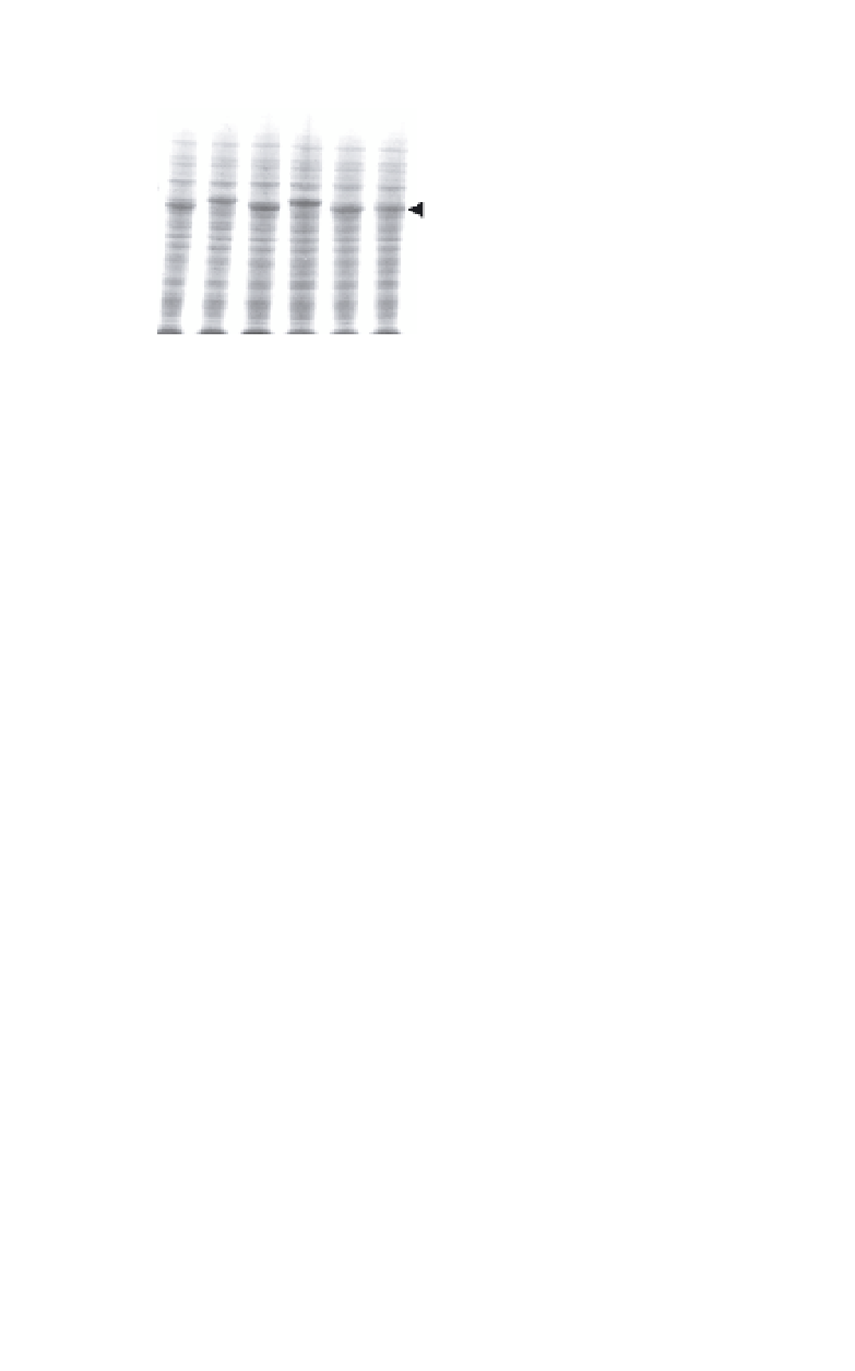
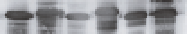
Fig. 2.
Expression of chimeric dORF2. A. Expression in the cell lysates was
examined by Coomasie brilliant blue staining. B. Antigenicity of the tag epi-
tope in the cell lysates was confirmed by Western blotting with the anti-tag
antibody. C. Presence of each chimeric dORF2 in cell supernatant (8
l) was
examined by Western blotting with the anti-tag antibody. The insertion site for
each chimera is indicated at the top of the panel. N; N-terminal, 1 to 4; sites
1 to 4, respectively, C; C-terminal. The arrowhead on the right of each panel
indicates the position of the chimeric dORF2.
µ
Western blot analysis (Fig. 2B). Even at the C-terminal region, where
the native HEV amino acid sequence is cleaved, the tag was not
cleaved off from dORF2 (Fig. 2B, lane C). Among these chimeras,
only the N- and C-terminal insertions resulted in release of a large
amount of chimeric dORF2 into the culture supernatant (Figs. 2C
and 2D), although small amounts were released when the insertions
were made at either site 3 or 4. These results indicate that internal
insertions somehow disturbed the release of dORF2 into the culture
supernatant. The precise mechanisms involved in the HEV virion for-
mation are not yet clear. The added tag at 52 amino acids upstream
from the C-terminal region, where dORF2 is normally cleaved in
insect cells, was not cleaved off in the infected cells during the gener-
ation of the chimeric VLPs. This is most likely due to alteration of the
amino acid sequence recognized by the proteolytic enzyme involved
in the C-terminal modification of HEV-VLP. The successful addition
of extra amino acid sequences to the C-terminal of dORF2 suggests
that the presence of extra amino acids at the C-terminal is not crucial