Biology Reference
In-Depth Information
A
B
MW
Anti-HEV
97.4
66
46
C
30
21.5
Anti-tag
CBB stain
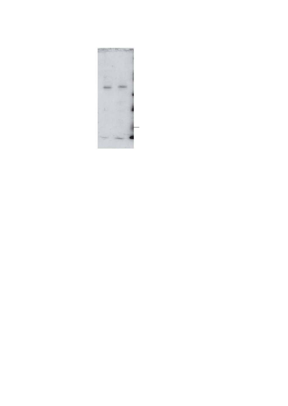
Fig. 3.
Purification of the chimeric VLP. Purified VLP-52C was analyzed
for its purity (A) and reactivity to anti-HEV (B) and anti-tag antibodies (C).
A. Equal amounts (0.3
52C) and VLP without
tag (w/o Tag) were separated on SDS-PAGE and stained by Coomasie bril-
liant blue staining. Positions of molecular weight markers are indicated on
the right of the panel. B and C. Equal amounts (0.1
µ
g) of purified VLP-52C (
−
52C)
and VLP without tag (w/o Tag) were analyzed by Western blotting using
anti-HEV (B) and anti-tag (C) antibodies, respectively.
µ
g) of VLP-52C (
−
for preventing dORF2 from being incorporated into a VLP form.
Rather, the amino acid sequences encoded by the HEV ORF2
genome prevented the formation of VLP.
We attempted to purify chimeric VLPs from the supernatant of
Tn5 cells expressing chimeric dORF2 with a tag at either C-termini.
The VLP-52C was slightly larger than the HEV-VLP without the tag
(Fig. 3A). The purified VLP-52C retained the antigenicity of HEV as
well as the intact tag epitope, as shown by the reactivity of specific
antibodies (Figs. 3B and 3C).
Electron microscopic observation showed that VLP-52C was
approximately 25 nm in diameter and indistinguishable from the