Biomedical Engineering Reference
In-Depth Information
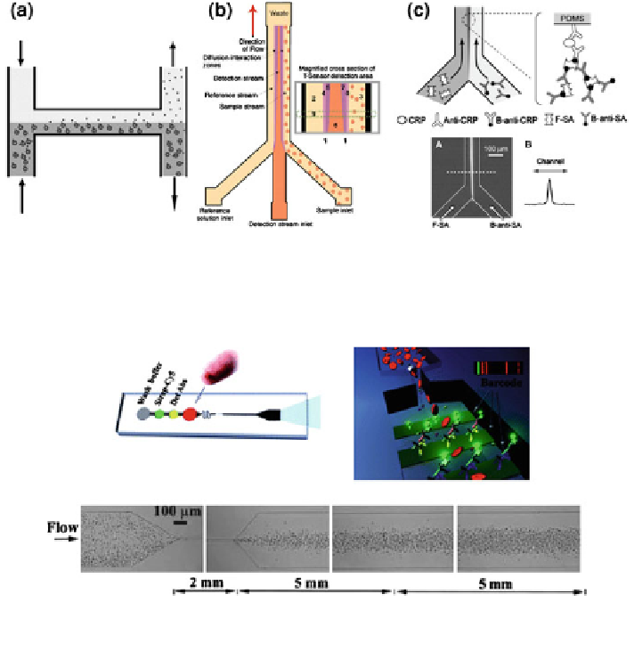
Fig. 3 Microfluidic separation by diffusion. a The H-filter Reprinted from [
90
]. b The T-sensor
Reprinted with permission from ref. [
113
]. c An immunoassay of human CRP based on the
T-sensor. Reprinted with permission from [
45
] Copyright (2007) American Chemical Society
Fig. 4 Analyte separation using the inertial forces generated by flow within microchannels.
Reproduced from [
110
] with permission of The Royal Society of Chemistry
enter a 500 lm wide chamber containing the captured antibodies via a narrow
10-20 lm microchannel. The fluid flow generated in this configuration produces
inertial lift forces that constrict the large cells to the centre-line of the chamber
(Fig.
4
). The theory behind this phenomenon has been studied in detail previously
[
13
,
122
]. The result is that the small target molecules can diffuse out into the
chamber and the cells do not interfere with the protein assay. The method was
successfully shown to extract and detect 11 proteins from whole blood.
Pretreatment can also be achieved using centrifugation microfluidic platforms.
These are typically fabricated on round structures which are spun at high speed to
generate centrifugal forces that drive the fluids through the channels. They are
often in the shape of compact discs, which makes them cheap and easy to operate
[
62
]. Moreover, their geometry allows for multiple parallel assays and easy inte-
gration with optical detectors [
61
]. There are a number of examples of microfluidic
centrifugation for separation applications [
37
,
40
,
55
,
100
]. A fully automated