Biomedical Engineering Reference
In-Depth Information
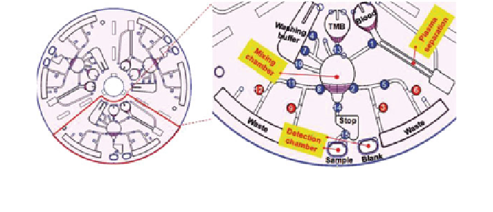
Fig. 5 On-chip centrifugation for pretreatment of whole blood. Reproduced from [
55
] with
permission of The Royal Society of Chemistry
device capable of detecting the antigen and antibody of the Hepatitis B virus
(HBV) was recently shown by Lee et al. [
55
]. After a whole blood sample is
introduced into the circular chip, it is inserted in a device and spun-up, separating
the plasma from the cells (Fig.
5
). Control of fluids was realised using valves
fabricated from ferromax (a mixture of paraffin wax and iron oxide nanoparticles),
which can be melted with a laser to allow the fluids to pass. Using optical
absorbance,
the
device
was
able
to
achieve
a
limit
of
detection
down
to
8.6 mIU mL
-1
in less than 30 min, making it applicable for POC use.
Microfluidic immunoassays. The high sensitivity and specificity of immuno-
assays makes them a powerful tool in molecular diagnostics. The current 'gold
standard' ELISA, while proven to be highly robust and reliable, suffers from large
sample requirements, long diffusion times, and hence, long incubation times [
97
].
Moreover, it requires a specific enzyme-fluorophore complex that does not inter-
fere with the reaction. In recent years, much research has focussed on combining
the classic ELISA approach with microfluidic technologies [
48
]. Such devices
enable quantitative immunoassay results by integrating sensitive optical, electrical,
or mechanical detection. The main advantages of a microfluidic immunoassay
compared to conventional approaches are: (i) fluidic control can be automated to
increase throughout and reproducibility (ii) lower consumption of precious
samples and expensive reagents and (iii) the increased surface-to-volume ratio
of the microchannels can speed up the antibody-antigen binding reactions. The
following section will highlight some of the more recent advances in microfluidic
immunoassays.
The microfluidic T-sensor, which can be exploited for use in immunoassays,
has been around since the late 1990s [
113
]. Like the H-filter, it takes advantage of
the laminar flow in microchannels and separates out the target antigen from
the sample based on diffusion (Fig.
3
b). The sample containing the antigen, the
antibody solution and a reference solution are all introduced separately into the
device where they meet in the main channel and begin to diffuse and mix [
113
].
Large molecules such as blood cells will not significantly diffuse into the other
streams; therefore, the smaller target molecules will move into the antibody stream