Biomedical Engineering Reference
In-Depth Information
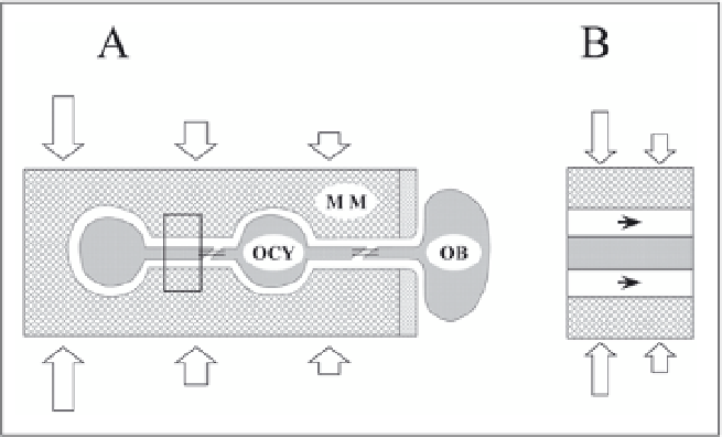
Figure 2. The concept of strain-derived canalicular flow. Deformation of the mineralized matrix (MM)
causes fluid pressure gradients within the canalicular system, resulting in fluid flow over the octeocyte
(OCY) processes. In response to the fluid shear stress osteocytes may signal the osteoblasts (OB) at the bone
surface.
Burger and Klein-Nulend
10
for a review). Comparison of the responsiveness of osteocytes,
osteoblasts and preosteoblasts to mechanical strain in vitro, showed that the osteocytes were
most responsive, more than osteoblasts and these more than preosteoblasts.
15-19
Thus, in the
course of differentiation from immature preosteoblast via osteoblast to osteocyte, bone cells
increase their sensitivity to mechanical strain. The manner whereby osteocytes sense the strains
of the mineralized matrix must be considered in light of the very small strains in bone during
daily loading, as compared to muscle tissue, for example.
20,21
This has led to the concept of
strain-derived canalicular fluid flow, or it's derivative fluid drag force, as the physical mediator
of mechano-sensing by osteocytes in bone tissue
20,22,23
(Fig. 2). Several experimental studies
support this concept. Animal studies show that bone formation in response to loading is sensi-
tive to strain rate, and that large strains alone are not sufficient to activate bone cells, suggesting
interstitial fluid flow as the physical stimulus involved.
24,25
In cell culture studies, fluid flow is
the most efficient cell activator,
26
and osteocytes show strong responses to fluid flow, including
rapid release of nitric oxide (NO) and prostaglandins.
15-19
Together these findings suggest that
the osteocyte network with it's accompanying lacuno-canalicular porosity, is the site of
mechanosensing in bone tissue. Mechanotransduction thus includes the translation, by osteo-
cytes, of canalicular flow into cell signals that can recruit osteoclasts and osteoblasts.
10
Mechanotransduction during Distraction Osteogenesis
Distraction osteogenesis (DOG) can be regarded as a form of bone engineering in vivo. It is
a well-established technique, initially used for lengthening of long bones,
5
but it has also found
other applications such as the treatment of limb deformities, bone defects and the creation of
craniofacial bone. The technique involves separation of a bone by osteotomy and fixation by an
external fixator. During a short latency period of about a week a collagenous connective tissue,
or callus, is formed. This newly formed soft tissue is then subjected to controlled distraction.
For appropriate distraction rates the tissue in the gap expands. Gradually ossification of the soft
tissue occurs and as a result the gap is bridged by new bone (Fig. 3). After consolidation, the
newly formed bone is remodeled as in existing bone.
Similar to fracture healing, soft tissue and bone formation in DOG is thought to be con-
trolled to a large part by growth factors, low-molecular weight glycoproteins. These factors are
released by the blood clot, the inflammatory cells that invade the fracture site and the bone
Search WWH ::

Custom Search