Biomedical Engineering Reference
In-Depth Information
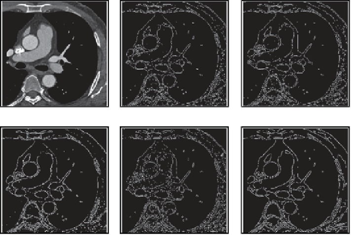
RULJLQDO
SUHZLWW
VREHO
UREHUWV
/R*
FDQQ\
Fig. 3.6
A DICOM slice image of a coronary artery in the axial plane is shown with different edge
detection algorithms applied
gradient based edge detection, the second order derivatives may be obtained by the
Laplacian of a Gaussian (LoG)
detector which applies a Laplacian of Gaussian
filter to look for zero crossings (Marr and Hildreth 1980); or the
Canny
edge detec-
tor (Canny 1986) which determines the local gradient maxima of the image based
on the derivative of a Gaussian filter. If two thresholds are used, then strong and
weak edges can be detected, which makes the method more robust in the presence
of noise. Each of these edge detectors are applied to the scanned coronary artery
used earlier (Fig.
3.6
).
Common problems arising from edge detection are primarily due to the absence
of an edge where a real border actually exists. In addition, the presence of noise,
fake, and weak edges will also have a negative influence on the algorithm. To over-
come this, detected edges are connected to build up the border into an edge chain
which will remove fake and weak edges. It should be noted that edge detection tech-
niques are typically used in conjunction with region-based technique for complete
segmentation.
3.3.4
Region Based Segmentation
In Sect. 3.3.2 regions were identified through threshold values based on the in-
tensity values of the pixels while in Sect. 3.3.3 the segmentation process involved
finding edge boundaries between regions based on pixel differences. In this section,
Search WWH ::

Custom Search