Biomedical Engineering Reference
In-Depth Information
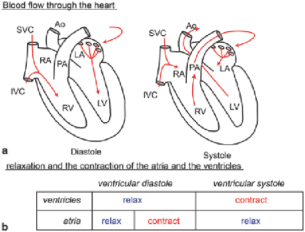
Fig. 2.6 a
Blood flow through the heart during ventricular diastole and systole (
adapted from
Klabunde (2005)
, cvphysiology.com).
b
Correspondence between the relaxation and the contrac-
tion of the atria and the ventricles with respect to the ventricular diastole and systole
atria and the ventricles go through these two stages every heartbeat, but the terms
diastole and systole alone, often refer to the ventricular stages. Figure
2.6
shows
how the blood travels through the heart during ventricular diastole and systole and
the correspondence between the relaxation and the contraction of the chambers with
respect to the ventricular phases.
2.2.3
Physiology of the Aorta
The aorta, which extends from the left ventricle in the upward direction and then
channels down towards the abdomen, is the largest and strongest artery in the human
body. Oxygenated blood is transported via this artery to the body organs through the
systemic circulation. Anatomically, the entire aorta is made up of three main seg-
ments: Ascending aorta; Aortic arch; and the Descending aorta (that comprises the
Thoracic aorta and the Abdominal aorta), labelled in Fig.
2.7
.
The aorta is a heterogeneous combination of smooth muscle, nerves, intimal
cells, endothelial cells, fibroblast-like cells, and a complex extracellular matrix. Its
wall is made up of several layers—the tunica adventitia, tunica media, and tunica
intima, which are mainly composed of collagen giving it stability by helping to an-
chor it to nearby organs. Once blood is squeezed out from the left ventricle, it trans-
ports the high pressure and pulsatile blood to the rest of the body. Being distensible
Search WWH ::

Custom Search