Biomedical Engineering Reference
In-Depth Information
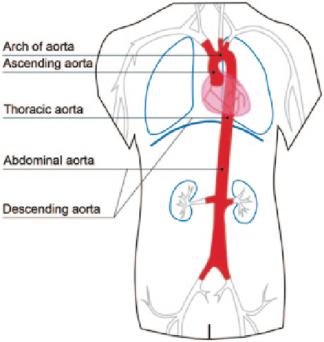
Fig. 2.7
Segments of the
aorta. The components of the
aorta are the ascending aorta,
aortic arch, and the descend-
ing aorta (
that comprises
the thoracic aorta, and the
abdominal aorta
). The aorta
is the largest and strongest
artery in the human body and
the main channel through
which blood is pumped from
the left ventricle. A network
of vessels is further branched
from the aorta to distribute
blood to the vital organs
and elastic, the blood pressure decreases in strength and becomes less pulsatile from
the aorta to arteries and to capillaries.
The blood spreads from the aorta down to the rest of the arteries which disperse
through the body in a branching pattern. This gives rise to the term the arterial tree
to describe the branching pattern of all the arteries in the body. The blood travels
through the arteries in a pulsatile manner. Reflected waves rebound at bifurcations
back to the semilunar valves and the aorta, which create a dicrotic notch in the
aortic pressure waveform when they push onto the aortic semilunar valve. This can
be visualised in the cardiac cycle profile (Fig.
2.8
) which shows a small dip that
coincides with the aortic valve closure. This dip is immediately followed by a short
rise, referred to as the dicrotic wave, then declines gradually.
As a body ages, the artery stiffens and causes the pulse wave to circulate faster
and the reflected waves return to the heart at a higher speed before the semilunar
valve closes, and resulting in higher blood pressure. Determining the pulse wave
velocity via invasive or non-invasive techniques can assess the arterial stiffness,
which is related to the degree of the disease.
2.2.4
Physiology of the Carotid Bifurcation
The carotid bifurcation, which includes the Common Carotid Artery (CCA), Exter-
nal Carotid Artery (ECA) and Internal Carotid Artery (ICA), transports oxygenated
Search WWH ::

Custom Search