Biomedical Engineering Reference
In-Depth Information
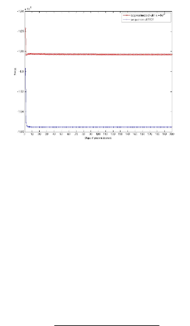
Fig. 4.
Energy in functional (6) of the solution obtained at each step of the gradient descent by
the approximated method for
=10
−
7
,
λ
=0
.
1
,
σ
=0
.
05
,
τ
=0
.
1
and by the new method for
λ
=0
.
1
,
σ
=0
.
05
,
τ
=0
.
1
Real Brain Images
Apart from the modelling exercise and the implementation details of the algorithm pre-
sented above, our main interest relies in the application of the proposed algorithm to
real brain images. In the following we present some preliminary results we are ob-
taining for Diffusion Weighted Magnetic Resonance Images (DW-MRI) denoising. The
DW-MR images are acquired and used for Diffusion Tensor Image (DTI) reconstruc-
tion, and the importance of the denoising step is crucial in DW-MRI analysis because
their characteristic very low SNR [6]. Diffusion Tensor Imaging is becoming one of the
most popular methods for the analysis of the white matter (WM) structure of the brain,
where some alterations can be found from early stages in some degenerative diseases.
This technique measures the Brownian motion (random motion) of the water molecules
in the brain, which is assumed to be isotropic when it is not restricted by the surround-
ing structures, since the WM regions contains densely packed fibre bundles they cause
an anisotropic diffusion of the water molecules along the perpendicular directions to
them. At each voxel of a DTI the water diffusion is represented by a symmetric
3
3
tensor, where the information of the preferred directions of the motion and the relevance
of these directions is found in the eigenvectors and the eigenvalues of the tensor. This
tensorial data can be represented as different scalar measurements, one of them is the
Fractional Anistropy (FA) of the tissue, which is defined as
×
3
(
λ
λ
3
)
2
λ
1
)
2
+(
λ
λ
2
)
2
+(
λ
−
−
−
FA
=
2(
λ
1
+
λ
2
+
λ
3
)


Search WWH ::

Custom Search